


 Foundational characteristics of cancer include proliferation, angiogenesis, migration, evasion of apoptosis, and cellular immortality. Find key markers for these cellular processes and antibodies to detect them.
 The SUMOplot™ Analysis Program predicts and scores sumoylation sites in your protein. SUMOylation is a post-translational modification involved in various cellular processes, such as nuclear-cytosolic transport, transcriptional regulation, apoptosis, protein stability, response to stress, and progression through the cell cycle.
 The Autophagy Receptor Motif Plotter predicts and scores autophagy receptor binding sites in your protein. Identifying proteins connected to this pathway is critical to understanding the role of autophagy in physiological as well as pathological processes such as development, differentiation, neurodegenerative diseases, stress, infection, and cancer.
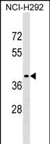
SNX21 Antibody (Center)
Affinity Purified Rabbit Polyclonal Antibody (Pab)
- SPECIFICATION
- CITATIONS
- PROTOCOLS
- BACKGROUND

Application
| WB, E |
|---|---|
| Primary Accession | Q969T3 |
| Other Accession | NP_219489.1 |
| Reactivity | Human |
| Host | Rabbit |
| Clonality | Polyclonal |
| Isotype | Rabbit IgG |
| Calculated MW | 41365 Da |
| Antigen Region | 210-236 aa |
| Gene ID | 90203 |
|---|---|
| Other Names | Sorting nexin-21, Sorting nexin L, SNX-L, SNX21, C20orf161, SNXL |
| Target/Specificity | This SNX21 antibody is generated from rabbits immunized with a KLH conjugated synthetic peptide between 210-236 amino acids from the Central region of human SNX21. |
| Dilution | WB~~1:1000 E~~Use at an assay dependent concentration. |
| Format | Purified polyclonal antibody supplied in PBS with 0.09% (W/V) sodium azide. This antibody is purified through a protein A column, followed by peptide affinity purification. |
| Storage | Maintain refrigerated at 2-8°C for up to 2 weeks. For long term storage store at -20°C in small aliquots to prevent freeze-thaw cycles. |
| Precautions | SNX21 Antibody (Center) is for research use only and not for use in diagnostic or therapeutic procedures. |
| Name | SNX21 |
|---|---|
| Synonyms | C20orf161, SNXL |
| Function | Binds to membranes enriched in phosphatidylinositol 3- phosphate (PtdIns(P3)) and phosphatidylinositol 4,5-bisphosphate. May be involved in several stages of intracellular trafficking. |
| Cellular Location | Cytoplasmic vesicle membrane {ECO:0000250|UniProtKB:Q3UR97}; Peripheral membrane protein; Cytoplasmic side. Early endosome membrane {ECO:0000250|UniProtKB:Q3UR97}; Peripheral membrane protein {ECO:0000250|UniProtKB:Q3UR97}; Cytoplasmic side {ECO:0000250|UniProtKB:Q3UR97} |
| Tissue Location | Highly expressed in fetus liver, but only weakly expressed in brain, skeleton muscle, smooth muscle, and cardiac muscle, kidney, and adrenal gland. |

Thousands of laboratories across the world have published research that depended on the performance of antibodies from Abcepta to advance their research. Check out links to articles that cite our products in major peer-reviewed journals, organized by research category.
info@abcepta.com, and receive a free "I Love Antibodies" mug.

Provided below are standard protocols that you may find useful for product applications.
Background
This gene encodes a member of the sorting nexin family. Members of this family contain a phox (PX) domain, which is a phosphoinositide binding domain, and are involved in intracellular trafficking. This protein does not contain a coiled coil region, like some family members. The specific function of this protein has not been determined. Multiple transcript variants encoding distinct isoforms have been identified for this gene.
References
Zeng, W., et al. Biochem. Biophys. Res. Commun. 299(4):542-548(2002)
Worby, C.A., et al. Nat. Rev. Mol. Cell Biol. 3(12):919-931(2002)
Deloukas, P., et al. Nature 414(6866):865-871(2001)
Xu, Y., et al. Biochem. J. 360 (PT 3), 513-530 (2001) :
Teasdale, R.D., et al. Biochem. J. 358 (PT 1), 7-16 (2001) :
If you have used an Abcepta product and would like to share how it has performed, please click on the "Submit Review" button and provide the requested information. Our staff will examine and post your review and contact you if needed.
If you have any additional inquiries please email technical services at tech@abcepta.com.
Ordering Information
Other Products
Shipping Information
