


 Foundational characteristics of cancer include proliferation, angiogenesis, migration, evasion of apoptosis, and cellular immortality. Find key markers for these cellular processes and antibodies to detect them.
 The SUMOplot™ Analysis Program predicts and scores sumoylation sites in your protein. SUMOylation is a post-translational modification involved in various cellular processes, such as nuclear-cytosolic transport, transcriptional regulation, apoptosis, protein stability, response to stress, and progression through the cell cycle.
 The Autophagy Receptor Motif Plotter predicts and scores autophagy receptor binding sites in your protein. Identifying proteins connected to this pathway is critical to understanding the role of autophagy in physiological as well as pathological processes such as development, differentiation, neurodegenerative diseases, stress, infection, and cancer.
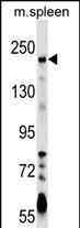
NEO1 Antibody (Center)
Affinity Purified Rabbit Polyclonal Antibody (Pab)
- SPECIFICATION
- CITATIONS
- PROTOCOLS
- BACKGROUND

Application
| WB, E |
|---|---|
| Primary Accession | Q92859 |
| Other Accession | P97603, P97798, NP_002490.2 |
| Reactivity | Human, Mouse |
| Predicted | Rat |
| Host | Rabbit |
| Clonality | Polyclonal |
| Isotype | Rabbit IgG |
| Calculated MW | 160017 Da |
| Antigen Region | 657-685 aa |
| Gene ID | 4756 |
|---|---|
| Other Names | Neogenin, Immunoglobulin superfamily DCC subclass member 2, NEO1, IGDCC2, NGN |
| Target/Specificity | This NEO1 antibody is generated from rabbits immunized with a KLH conjugated synthetic peptide between 657-685 amino acids from the Central region of human NEO1. |
| Dilution | WB~~1:1000 E~~Use at an assay dependent concentration. |
| Format | Purified polyclonal antibody supplied in PBS with 0.09% (W/V) sodium azide. This antibody is purified through a protein A column, followed by peptide affinity purification. |
| Storage | Maintain refrigerated at 2-8°C for up to 2 weeks. For long term storage store at -20°C in small aliquots to prevent freeze-thaw cycles. |
| Precautions | NEO1 Antibody (Center) is for research use only and not for use in diagnostic or therapeutic procedures. |
| Name | NEO1 |
|---|---|
| Synonyms | IGDCC2, NGN |
| Function | Multi-functional cell surface receptor regulating cell adhesion in many diverse developmental processes, including neural tube and mammary gland formation, myogenesis and angiogenesis. Receptor for members of the BMP, netrin, and repulsive guidance molecule (RGM) families. Netrin-Neogenin interactions result in a chemoattractive axon guidance response and cell-cell adhesion, the interaction between NEO1/Neogenin and RGMa and RGMb induces a chemorepulsive response. |
| Cellular Location | Cell membrane; Single-pass type I membrane protein |
| Tissue Location | Widely expressed and also in cancer cell lines. |

Thousands of laboratories across the world have published research that depended on the performance of antibodies from Abcepta to advance their research. Check out links to articles that cite our products in major peer-reviewed journals, organized by research category.
info@abcepta.com, and receive a free "I Love Antibodies" mug.

Provided below are standard protocols that you may find useful for product applications.
Background
This gene encodes a cell surface protein that is a member of the immunoglobulin superfamily. The encoded protein consists of four N-terminal immunoglobulin-like domains, six fibronectin type III domains, a transmembrane domain and a C-terminal internal domain that shares homology with the tumor suppressor candidate gene DCC. This protein may be involved in cell growth and differentiation and in cell-cell adhesion. Defects in this gene are associated with cell proliferation in certain cancers. Alternate splicing results in multiple transcript variants. [provided by RefSeq].
References
Bradford, D., et al. J. Comp. Neurol. 518(16):3237-3253(2010)
Davila, S., et al. Genes Immun. 11(3):232-238(2010)
Zhang, A.S., et al. J. Biol. Chem. 284(34):22580-22589(2009)
Fujita, Y., et al. Cell Death Differ. 15(10):1593-1608(2008)
Zhang, A.S., et al. J. Biol. Chem. 283(25):17494-17502(2008)
If you have used an Abcepta product and would like to share how it has performed, please click on the "Submit Review" button and provide the requested information. Our staff will examine and post your review and contact you if needed.
If you have any additional inquiries please email technical services at tech@abcepta.com.
Ordering Information
Shipping Information
