


 Foundational characteristics of cancer include proliferation, angiogenesis, migration, evasion of apoptosis, and cellular immortality. Find key markers for these cellular processes and antibodies to detect them.
 The SUMOplot™ Analysis Program predicts and scores sumoylation sites in your protein. SUMOylation is a post-translational modification involved in various cellular processes, such as nuclear-cytosolic transport, transcriptional regulation, apoptosis, protein stability, response to stress, and progression through the cell cycle.
 The Autophagy Receptor Motif Plotter predicts and scores autophagy receptor binding sites in your protein. Identifying proteins connected to this pathway is critical to understanding the role of autophagy in physiological as well as pathological processes such as development, differentiation, neurodegenerative diseases, stress, infection, and cancer.
DHRS1 Antibody (Center)
Affinity Purified Rabbit Polyclonal Antibody (Pab)
- SPECIFICATION
- CITATIONS
- PROTOCOLS
- BACKGROUND

Application
| WB, E |
|---|---|
| Primary Accession | Q96LJ7 |
| Other Accession | NP_001129522.1, NP_612461.1 |
| Reactivity | Human |
| Host | Rabbit |
| Clonality | Polyclonal |
| Isotype | Rabbit IgG |
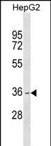
| Calculated MW | 33909 Da |
| Antigen Region | 183-209 aa |
| Gene ID | 115817 |
|---|---|
| Other Names | Dehydrogenase/reductase SDR family member 1, 11--, DHRS1 |
| Target/Specificity | This DHRS1 antibody is generated from rabbits immunized with a KLH conjugated synthetic peptide between 183-209 amino acids from the Central region of human DHRS1. |
| Dilution | WB~~1:1000 E~~Use at an assay dependent concentration. |
| Format | Purified polyclonal antibody supplied in PBS with 0.09% (W/V) sodium azide. This antibody is purified through a protein A column, followed by peptide affinity purification. |
| Storage | Maintain refrigerated at 2-8°C for up to 2 weeks. For long term storage store at -20°C in small aliquots to prevent freeze-thaw cycles. |
| Precautions | DHRS1 Antibody (Center) is for research use only and not for use in diagnostic or therapeutic procedures. |
| Name | DHRS1 (HGNC:16445) |
|---|---|
| Synonyms | SDR19C1 |
| Function | NADPH-dependent oxidoreductase which catalyzes the reduction of steroids (estrone, androstene-3,17-dione and cortisone) as well as prostaglandin E1, isatin and xenobiotics in vitro (PubMed:30031147). May have a role in steroid and/or xenobiotic metabolism (PubMed:30031147). |
| Cellular Location | Endoplasmic reticulum. Note=May be attached to the ER membrane by its C-terminus segment. |
| Tissue Location | Detected in heart, liver, adrenal glands, and at low levels in skeletal muscle, kidney, pancreas and brain |

Thousands of laboratories across the world have published research that depended on the performance of antibodies from Abcepta to advance their research. Check out links to articles that cite our products in major peer-reviewed journals, organized by research category.
info@abcepta.com, and receive a free "I Love Antibodies" mug.

Provided below are standard protocols that you may find useful for product applications.
Background
This gene encodes a member of the short-chain dehydrogenases/reductases (SDR) family. The encoded enzyme contains a conserved catalytic domain and likely functions as an oxidoreductase. Multiple alternatively spliced variants, encoding the same protein, have been identified.
References
Persson, B., et al. Chem. Biol. Interact. 178 (1-3), 94-98 (2009) :
Wu, Q., et al. Mol. Biol. Rep. 28(4):193-198(2001)
If you have used an Abcepta product and would like to share how it has performed, please click on the "Submit Review" button and provide the requested information. Our staff will examine and post your review and contact you if needed.
If you have any additional inquiries please email technical services at tech@abcepta.com.
Ordering Information
Other Products
Shipping Information
