


 Foundational characteristics of cancer include proliferation, angiogenesis, migration, evasion of apoptosis, and cellular immortality. Find key markers for these cellular processes and antibodies to detect them.
 The SUMOplot™ Analysis Program predicts and scores sumoylation sites in your protein. SUMOylation is a post-translational modification involved in various cellular processes, such as nuclear-cytosolic transport, transcriptional regulation, apoptosis, protein stability, response to stress, and progression through the cell cycle.
 The Autophagy Receptor Motif Plotter predicts and scores autophagy receptor binding sites in your protein. Identifying proteins connected to this pathway is critical to understanding the role of autophagy in physiological as well as pathological processes such as development, differentiation, neurodegenerative diseases, stress, infection, and cancer.
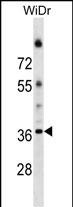
ALG5 Antibody (Center)
Affinity Purified Rabbit Polyclonal Antibody (Pab)
- SPECIFICATION
- CITATIONS
- PROTOCOLS
- BACKGROUND

Application
| WB, E |
|---|---|
| Primary Accession | Q9Y673 |
| Other Accession | NP_037470.1 |
| Reactivity | Human |
| Host | Rabbit |
| Clonality | Polyclonal |
| Isotype | Rabbit IgG |
| Calculated MW | 36946 Da |
| Antigen Region | 74-100 aa |
| Gene ID | 29880 |
|---|---|
| Other Names | Dolichyl-phosphate beta-glucosyltransferase, DolP-glucosyltransferase, Asparagine-linked glycosylation protein 5 homolog, ALG5 |
| Target/Specificity | This ALG5 antibody is generated from rabbits immunized with a KLH conjugated synthetic peptide between 74-100 amino acids from the Central region of human ALG5. |
| Dilution | WB~~1:1000 E~~Use at an assay dependent concentration. |
| Format | Purified polyclonal antibody supplied in PBS with 0.09% (W/V) sodium azide. This antibody is purified through a protein A column, followed by peptide affinity purification. |
| Storage | Maintain refrigerated at 2-8°C for up to 2 weeks. For long term storage store at -20°C in small aliquots to prevent freeze-thaw cycles. |
| Precautions | ALG5 Antibody (Center) is for research use only and not for use in diagnostic or therapeutic procedures. |
| Name | ALG5 (HGNC:20266) |
|---|---|
| Function | Dolichyl-phosphate beta-glucosyltransferase that operates in the biosynthetic pathway of dolichol-linked oligosaccharides, the glycan precursors employed in protein asparagine (N)-glycosylation. The assembly of dolichol-linked oligosaccharides begins on the cytosolic side of the endoplasmic reticulum membrane and finishes in its lumen. The sequential addition of sugars to dolichol pyrophosphate produces dolichol-linked oligosaccharides containing fourteen sugars, including two GlcNAcs, nine mannoses and three glucoses. Once assembled, the oligosaccharide is transferred from the lipid to nascent proteins by oligosaccharyltransferases. Dolichyl-phosphate beta-glucosyltransferase produces dolichyl beta-D-glucosyl phosphate/Dol-P-Glc, the glucose donor substrate used sequentially by ALG6, ALG8 and ALG10 to add glucose residues on top of the Man(9)GlcNAc(2)-PP-Dol structure. These are the three last steps in the biosynthetic pathway of dolichol-linked oligosaccharides to produce Glc(3)Man(9)GlcNAc(2)-PP-Dol. The enzyme is most probably active on the cytoplasmic side of the endoplasmic reticulum while its product Dol-P-Glc is the substrate for ALG6, ALG8 and ALG11 in the lumen of the endoplasmic reticulum. |
| Cellular Location | Endoplasmic reticulum membrane; Single-pass membrane protein |
| Tissue Location | Expressed in pancreas, placenta, liver, heart, brain, kidney, skeletal muscle, and lung |

Thousands of laboratories across the world have published research that depended on the performance of antibodies from Abcepta to advance their research. Check out links to articles that cite our products in major peer-reviewed journals, organized by research category.
info@abcepta.com, and receive a free "I Love Antibodies" mug.

Provided below are standard protocols that you may find useful for product applications.
Background
This gene encodes a member of the glycosyltransferase 2 family. The encoded protein participates in glucosylation of the oligomannose core in N-linked glycosylation of proteins. The addition of glucose residues to the oligomannose core is necessary to ensure substrate recognition, and therefore, effectual transfer of the oligomannose core to the nascent glycoproteins. Multiple transcript variants encoding different isoforms have been found for this gene.
References
Imbach, T., et al. Proc. Natl. Acad. Sci. U.S.A. 96(12):6982-6987(1999)
If you have used an Abcepta product and would like to share how it has performed, please click on the "Submit Review" button and provide the requested information. Our staff will examine and post your review and contact you if needed.
If you have any additional inquiries please email technical services at tech@abcepta.com.
Ordering Information
Other Products
Shipping Information
