


 Foundational characteristics of cancer include proliferation, angiogenesis, migration, evasion of apoptosis, and cellular immortality. Find key markers for these cellular processes and antibodies to detect them.
 The SUMOplot™ Analysis Program predicts and scores sumoylation sites in your protein. SUMOylation is a post-translational modification involved in various cellular processes, such as nuclear-cytosolic transport, transcriptional regulation, apoptosis, protein stability, response to stress, and progression through the cell cycle.
 The Autophagy Receptor Motif Plotter predicts and scores autophagy receptor binding sites in your protein. Identifying proteins connected to this pathway is critical to understanding the role of autophagy in physiological as well as pathological processes such as development, differentiation, neurodegenerative diseases, stress, infection, and cancer.
FIGLA Antibody (Center)
Affinity Purified Rabbit Polyclonal Antibody (Pab)
- SPECIFICATION
- CITATIONS
- PROTOCOLS
- BACKGROUND

Application
| WB, E |
|---|---|
| Primary Accession | Q6QHK4 |
| Other Accession | NP_001004311.2 |
| Reactivity | Human |
| Host | Rabbit |
| Clonality | Polyclonal |
| Isotype | Rabbit IgG |
| Calculated MW | 24123 Da |
| Antigen Region | 108-137 aa |
| Gene ID | 344018 |
|---|---|
| Other Names | Factor in the germline alpha, FIGalpha, Class C basic helix-loop-helix protein 8, bHLHc8, Folliculogenesis-specific basic helix-loop-helix protein, Transcription factor FIGa, FIGLA, BHLHC8 |
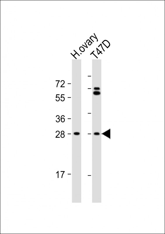
| Target/Specificity | This FIGLA antibody is generated from rabbits immunized with a KLH conjugated synthetic peptide between 108-137 amino acids from the Central region of human FIGLA. |
| Dilution | WB~~1:1000 E~~Use at an assay dependent concentration. |
| Format | Purified polyclonal antibody supplied in PBS with 0.09% (W/V) sodium azide. This antibody is purified through a protein A column, followed by peptide affinity purification. |
| Storage | Maintain refrigerated at 2-8°C for up to 2 weeks. For long term storage store at -20°C in small aliquots to prevent freeze-thaw cycles. |
| Precautions | FIGLA Antibody (Center) is for research use only and not for use in diagnostic or therapeutic procedures. |
| Name | FIGLA |
|---|---|
| Synonyms | BHLHC8 |
| Function | Germline specific transcription factor implicated in postnatal oocyte-specific gene expression. Plays a key regulatory role in the expression of multiple oocyte-specific genes, including those that initiate folliculogenesis and those that encode the zona pellucida (ZP1, ZP2 and ZP3) required for fertilization and early embryonic survival. Essential for oocytes to survive and form primordial follicles. The persistence of FIGLA in adult females suggests that it may regulate additional pathways that are essential for normal ovarian development. Binds to the E-box (5'-CANNTG-3') of the ZPs (ZP1, ZP2, ZP3) promoters. |
| Cellular Location | Nucleus. |
| Tissue Location | Germ cells. Expressed in the fetal ovary, but not by a range of other tissues. Expression increases across mid-gestation, rising some 40-fold by the time of primordial follicle formation |

Thousands of laboratories across the world have published research that depended on the performance of antibodies from Abcepta to advance their research. Check out links to articles that cite our products in major peer-reviewed journals, organized by research category.
info@abcepta.com, and receive a free "I Love Antibodies" mug.

Provided below are standard protocols that you may find useful for product applications.
Background
This gene encodes a protein that functions in postnatal oocyte-specific gene expression. The protein is a basic helix-loop-helix transcription factor that regulates multiple oocyte-specific genes, including genes involved in folliculogenesis and those that encode the zona pellucida. Mutations in this gene cause premature ovarian failure type 6.
References
van Dooren, M.F., et al. Curr. Opin. Obstet. Gynecol. 21(4):313-317(2009)
Fowler, P.A., et al. J. Clin. Endocrinol. Metab. 94(4):1427-1435(2009)
Tormala, R.M., et al. Mol. Cell. Endocrinol. 289 (1-2), 10-15 (2008) :
Zhao, H., et al. Am. J. Hum. Genet. 82(6):1342-1348(2008)
Suzumori, N., et al. Curr. Med. Chem. 14(3):353-357(2007)
If you have used an Abcepta product and would like to share how it has performed, please click on the "Submit Review" button and provide the requested information. Our staff will examine and post your review and contact you if needed.
If you have any additional inquiries please email technical services at tech@abcepta.com.
Ordering Information
Other Products
Shipping Information
