


 Foundational characteristics of cancer include proliferation, angiogenesis, migration, evasion of apoptosis, and cellular immortality. Find key markers for these cellular processes and antibodies to detect them.
 The SUMOplot™ Analysis Program predicts and scores sumoylation sites in your protein. SUMOylation is a post-translational modification involved in various cellular processes, such as nuclear-cytosolic transport, transcriptional regulation, apoptosis, protein stability, response to stress, and progression through the cell cycle.
 The Autophagy Receptor Motif Plotter predicts and scores autophagy receptor binding sites in your protein. Identifying proteins connected to this pathway is critical to understanding the role of autophagy in physiological as well as pathological processes such as development, differentiation, neurodegenerative diseases, stress, infection, and cancer.
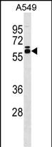
DUS2L Antibody (C-term)
Affinity Purified Rabbit Polyclonal Antibody (Pab)
- SPECIFICATION
- CITATIONS
- PROTOCOLS
- BACKGROUND

Application
| WB, E |
|---|---|
| Primary Accession | Q9NX74 |
| Other Accession | NP_060273.1 |
| Reactivity | Human |
| Host | Rabbit |
| Clonality | Polyclonal |
| Isotype | Rabbit IgG |
| Calculated MW | 55050 Da |
| Antigen Region | 371-397 aa |
| Gene ID | 54920 |
|---|---|
| Other Names | tRNA-dihydrouridine(20) synthase [NAD(P)+]-like, 131-, Dihydrouridine synthase 2, Up-regulated in lung cancer protein 8, URLC8, tRNA-dihydrouridine synthase 2-like, hDUS2, DUS2, DUS2L |
| Target/Specificity | This DUS2L antibody is generated from rabbits immunized with a KLH conjugated synthetic peptide between 371-397 amino acids from the C-terminal region of human DUS2L. |
| Dilution | WB~~1:1000 E~~Use at an assay dependent concentration. |
| Format | Purified polyclonal antibody supplied in PBS with 0.09% (W/V) sodium azide. This antibody is purified through a protein A column, followed by peptide affinity purification. |
| Storage | Maintain refrigerated at 2-8°C for up to 2 weeks. For long term storage store at -20°C in small aliquots to prevent freeze-thaw cycles. |
| Precautions | DUS2L Antibody (C-term) is for research use only and not for use in diagnostic or therapeutic procedures. |
| Name | DUS2 |
|---|---|
| Synonyms | DUS2L |
| Function | Catalyzes the NADPH-dependent synthesis of dihydrouridine, a modified base found in the D-loop of most tRNAs (PubMed:15994936, PubMed:26429968, PubMed:30149704, PubMed:34798057, PubMed:38680565). Specifically modifies U20 in cytoplasmic tRNAs (PubMed:38680565). Activity depends on the presence of guanosine at position 19 in the tRNA substrate (PubMed:38680565). Negatively regulates the activation of EIF2AK2/PKR (PubMed:18096616). |
| Cellular Location | Cytoplasm. Endoplasmic reticulum. Note=Mainly at the endoplasmic reticulum. |
| Tissue Location | Weak expression in heart, placenta and skeletal muscle. Up-regulated in most lung cancer cells (at protein level) |

Thousands of laboratories across the world have published research that depended on the performance of antibodies from Abcepta to advance their research. Check out links to articles that cite our products in major peer-reviewed journals, organized by research category.
info@abcepta.com, and receive a free "I Love Antibodies" mug.

Provided below are standard protocols that you may find useful for product applications.
Background
Dihydrouridine synthase catalyzes reduction of the 5,6-double bond of a uridine residue on the displacement loop of tRNA. The resultant modified base, 5,6-dihydrouridine, appears to increase the conformational flexibility and dynamic motion of tRNA (Kato et al., 2005 [PubMed 15994936]).
References
Mittelstadt, M., et al. Nucleic Acids Res. 36(3):998-1008(2008)
Sugiyama, N., et al. Mol. Cell Proteomics 6(6):1103-1109(2007)
Lamesch, P., et al. Genomics 89(3):307-315(2007)
Kato, T., et al. Cancer Res. 65(13):5638-5646(2005)
If you have used an Abcepta product and would like to share how it has performed, please click on the "Submit Review" button and provide the requested information. Our staff will examine and post your review and contact you if needed.
If you have any additional inquiries please email technical services at tech@abcepta.com.
Ordering Information
Other Products
Shipping Information
