


 Foundational characteristics of cancer include proliferation, angiogenesis, migration, evasion of apoptosis, and cellular immortality. Find key markers for these cellular processes and antibodies to detect them.
 The SUMOplot™ Analysis Program predicts and scores sumoylation sites in your protein. SUMOylation is a post-translational modification involved in various cellular processes, such as nuclear-cytosolic transport, transcriptional regulation, apoptosis, protein stability, response to stress, and progression through the cell cycle.
 The Autophagy Receptor Motif Plotter predicts and scores autophagy receptor binding sites in your protein. Identifying proteins connected to this pathway is critical to understanding the role of autophagy in physiological as well as pathological processes such as development, differentiation, neurodegenerative diseases, stress, infection, and cancer.
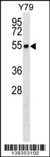
RNF26 Antibody (Center)
Affinity Purified Rabbit Polyclonal Antibody (Pab)
- SPECIFICATION
- CITATIONS
- PROTOCOLS
- BACKGROUND

Application
| WB, E |
|---|---|
| Primary Accession | Q9BY78 |
| Other Accession | NP_114404.1 |
| Reactivity | Human |
| Host | Rabbit |
| Clonality | Polyclonal |
| Isotype | Rabbit IgG |
| Calculated MW | 47737 Da |
| Antigen Region | 247-273 aa |
| Gene ID | 79102 |
|---|---|
| Other Names | RING finger protein 26, RNF26 |
| Target/Specificity | This RNF26 antibody is generated from rabbits immunized with a KLH conjugated synthetic peptide between 247-273 amino acids from the Central region of human RNF26. |
| Dilution | WB~~1:1000 E~~Use at an assay dependent concentration. |
| Format | Purified polyclonal antibody supplied in PBS with 0.09% (W/V) sodium azide. This antibody is purified through a protein A column, followed by peptide affinity purification. |
| Storage | Maintain refrigerated at 2-8°C for up to 2 weeks. For long term storage store at -20°C in small aliquots to prevent freeze-thaw cycles. |
| Precautions | RNF26 Antibody (Center) is for research use only and not for use in diagnostic or therapeutic procedures. |
| Name | RNF26 (HGNC:14646) |
|---|---|
| Function | E3 ubiquitin-protein ligase that plays a key role in endosome organization by retaining vesicles in the perinuclear cloud (PubMed:27368102). Acts as a platform for perinuclear positioning of the endosomal system by mediating ubiquitination of SQSTM1 through interaction with the ubiquitin conjugating enzyme UBE2J1 (PubMed:27368102, PubMed:33472082). Ubiquitinated SQSTM1 attracts specific vesicle-associated adapters, forming a molecular bridge that restrains cognate vesicles in the perinuclear region and organizes the endosomal pathway for efficient cargo transport (PubMed:27368102, PubMed:33472082). Also acts as a regulator of type I interferon production in response to viral infection by mediating the formation of 'Lys-11'-linked polyubiquitin chains on TMEM173/STING, leading to stabilize TMEM173/STING (PubMed:25254379, PubMed:32614325). Also required to limit type I interferon response by promoting autophagic degradation of IRF3 (PubMed:25254379). |
| Cellular Location | Endoplasmic reticulum membrane; Multi-pass membrane protein |
| Tissue Location | Ubiquitous. Up-regulated in several cancer cell lines. |

Thousands of laboratories across the world have published research that depended on the performance of antibodies from Abcepta to advance their research. Check out links to articles that cite our products in major peer-reviewed journals, organized by research category.
info@abcepta.com, and receive a free "I Love Antibodies" mug.

Provided below are standard protocols that you may find useful for product applications.
Background
The protein encoded by this intronless gene contains a C3HC5 type of RING finger, a motif known to be involved in protein-DNA and protein-protein interactions. The expression of this gene was found to be upregulated in cancer cell lines derived from different types of cancer.
References
Bailey, S.D., et al. Diabetes Care (2010) In press :
Talmud, P.J., et al. Am. J. Hum. Genet. 85(5):628-642(2009)
Katoh, M. Biochem. Biophys. Res. Commun. 282(4):1038-1044(2001)
If you have used an Abcepta product and would like to share how it has performed, please click on the "Submit Review" button and provide the requested information. Our staff will examine and post your review and contact you if needed.
If you have any additional inquiries please email technical services at tech@abcepta.com.
Ordering Information
Other Products
Shipping Information
