


 Foundational characteristics of cancer include proliferation, angiogenesis, migration, evasion of apoptosis, and cellular immortality. Find key markers for these cellular processes and antibodies to detect them.
 The SUMOplot™ Analysis Program predicts and scores sumoylation sites in your protein. SUMOylation is a post-translational modification involved in various cellular processes, such as nuclear-cytosolic transport, transcriptional regulation, apoptosis, protein stability, response to stress, and progression through the cell cycle.
 The Autophagy Receptor Motif Plotter predicts and scores autophagy receptor binding sites in your protein. Identifying proteins connected to this pathway is critical to understanding the role of autophagy in physiological as well as pathological processes such as development, differentiation, neurodegenerative diseases, stress, infection, and cancer.
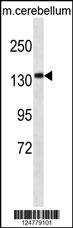
CAMSAP3 Antibody (N-term)
Affinity Purified Rabbit Polyclonal Antibody (Pab)
- SPECIFICATION
- CITATIONS: 1
- PROTOCOLS
- BACKGROUND

Application
| WB, E |
|---|---|
| Primary Accession | Q9P1Y5 |
| Other Accession | NP_065953.1 |
| Reactivity | Human, Mouse |
| Host | Rabbit |
| Clonality | Polyclonal |
| Isotype | Rabbit IgG |
| Calculated MW | 134750 Da |
| Antigen Region | 103-131 aa |
| Gene ID | 57662 |
|---|---|
| Other Names | Calmodulin-regulated spectrin-associated protein 3, Protein Nezha, CAMSAP3, KIAA1543 |
| Target/Specificity | This CAMSAP3 antibody is generated from rabbits immunized with a KLH conjugated synthetic peptide between 103-131 amino acids from the N-terminal region of human CAMSAP3. |
| Dilution | WB~~1:1000 E~~Use at an assay dependent concentration. |
| Format | Purified polyclonal antibody supplied in PBS with 0.09% (W/V) sodium azide. This antibody is purified through a protein A column, followed by peptide affinity purification. |
| Storage | Maintain refrigerated at 2-8°C for up to 2 weeks. For long term storage store at -20°C in small aliquots to prevent freeze-thaw cycles. |
| Precautions | CAMSAP3 Antibody (N-term) is for research use only and not for use in diagnostic or therapeutic procedures. |
| Name | CAMSAP3 (HGNC:29307) |
|---|---|
| Function | Key microtubule-organizing protein that specifically binds the minus-end of non-centrosomal microtubules and regulates their dynamics and organization (PubMed:19041755, PubMed:23169647). Specifically recognizes growing microtubule minus-ends and autonomously decorates and stabilizes microtubule lattice formed by microtubule minus-end polymerization (PubMed:24486153). Acts on free microtubule minus-ends that are not capped by microtubule-nucleating proteins or other factors and protects microtubule minus-ends from depolymerization (PubMed:24486153). In addition, it also reduces the velocity of microtubule polymerization (PubMed:24486153). Required for the biogenesis and the maintenance of zonula adherens by anchoring the minus-end of microtubules to zonula adherens and by recruiting the kinesin KIFC3 to those junctional sites (PubMed:19041755). Required for orienting the apical-to-basal polarity of microtubules in epithelial cells: acts by tethering non-centrosomal microtubules to the apical cortex, leading to their longitudinal orientation (PubMed:26715742, PubMed:27802168). Plays a key role in early embryos, which lack centrosomes: accumulates at the microtubule bridges that connect pairs of cells and enables the formation of a non-centrosomal microtubule- organizing center that directs intracellular transport in the early embryo (By similarity). Couples non-centrosomal microtubules with actin: interaction with MACF1 at the minus ends of non-centrosomal microtubules, tethers the microtubules to actin filaments, regulating focal adhesion size and cell migration (PubMed:27693509). Plays a key role in the generation of non-centrosomal microtubules by accumulating in the pericentrosomal region and cooperating with KATNA1 to release non-centrosomal microtubules from the centrosome (PubMed:28386021). Through the microtubule cytoskeleton, also regulates the organization of cellular organelles including the Golgi and the early endosomes (PubMed:28089391). Through interaction with AKAP9, involved in translocation of Golgi vesicles in epithelial cells, where microtubules are mainly non-centrosomal (PubMed:28089391). Plays an important role in motile cilia function by facilitatating proper orientation of basal bodies and formation of central microtubule pairs in motile cilia (By similarity). |
| Cellular Location | Cytoplasm, cytoskeleton. Cell junction, adherens junction. Cytoplasm Cytoplasm, cytoskeleton, cilium axoneme {ECO:0000250|UniProtKB:Q80VC9} Cytoplasm, cytoskeleton, cilium basal body {ECO:0000250|UniProtKB:Q80VC9}. Note=Scattered in the cytoplasm, associated with the minus-end of microtubules and also detected at the centrosomes (PubMed:19041755, PubMed:24486153, PubMed:27693509) Decorates the minus-end of microtubules by decreasing the rate of tubulin incorporation and remaining bound (PubMed:24486153). Localizes along zonula adherens only at mature cell-cell contacts (PubMed:19041755). In early embryos, accumulates at the microtubule bridges that connect pairs of cells: this structure is present in early embryos, which lack centrosomes (By similarity). This cytokinetic bridge does not undergo stereotypical abscission after cell division (By similarity). Accumulates to the pericentrosomal region following interaction with KATNA1 (PubMed:28386021) {ECO:0000250|UniProtKB:Q80VC9, ECO:0000269|PubMed:19041755, ECO:0000269|PubMed:24486153, ECO:0000269|PubMed:27693509, ECO:0000269|PubMed:28386021} |

Provided below are standard protocols that you may find useful for product applications.
Background
Microtubule minus-end binding protein that acts as a regulator of microtubule dynamics. Specifically required for zonula adherens biogenesis and maintenance by anchoring microtubules at their minus-ends to zonula adherens, leading to recruit KIFC3 kinesin to junctional site.
References
Meng, W., et al. Cell 135(5):948-959(2008)
Akhmanova, A., et al. Cell 135(5):791-793(2008)
Olsen, J.V., et al. Cell 127(3):635-648(2006)
Beausoleil, S.A., et al. Nat. Biotechnol. 24(10):1285-1292(2006)
Beausoleil, S.A., et al. Proc. Natl. Acad. Sci. U.S.A. 101(33):12130-12135(2004)
If you have used an Abcepta product and would like to share how it has performed, please click on the "Submit Review" button and provide the requested information. Our staff will examine and post your review and contact you if needed.
If you have any additional inquiries please email technical services at tech@abcepta.com.
Ordering Information
Other Products
Shipping Information
