


 Foundational characteristics of cancer include proliferation, angiogenesis, migration, evasion of apoptosis, and cellular immortality. Find key markers for these cellular processes and antibodies to detect them.
 The SUMOplot™ Analysis Program predicts and scores sumoylation sites in your protein. SUMOylation is a post-translational modification involved in various cellular processes, such as nuclear-cytosolic transport, transcriptional regulation, apoptosis, protein stability, response to stress, and progression through the cell cycle.
 The Autophagy Receptor Motif Plotter predicts and scores autophagy receptor binding sites in your protein. Identifying proteins connected to this pathway is critical to understanding the role of autophagy in physiological as well as pathological processes such as development, differentiation, neurodegenerative diseases, stress, infection, and cancer.
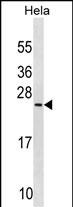
PRELID1 Antibody (N-term)
Affinity Purified Rabbit Polyclonal Antibody (Pab)
- SPECIFICATION
- CITATIONS
- PROTOCOLS
- BACKGROUND

Application
| WB, E |
|---|---|
| Primary Accession | Q9Y255 |
| Other Accession | Q8R107, NP_037369.1 |
| Reactivity | Human |
| Predicted | Mouse |
| Host | Rabbit |
| Clonality | Polyclonal |
| Isotype | Rabbit IgG |
| Calculated MW | 25181 Da |
| Antigen Region | 27-54 aa |
| Gene ID | 27166 |
|---|---|
| Other Names | PRELI domain-containing protein 1, mitochondrial, 25 kDa protein of relevant evolutionary and lymphoid interest, Px19-like protein, PRELID1, PRELI |
| Target/Specificity | This PRELID1 antibody is generated from rabbits immunized with a KLH conjugated synthetic peptide between 27-54 amino acids from the N-terminal region of human PRELID1. |
| Dilution | WB~~1:1000 E~~Use at an assay dependent concentration. |
| Format | Purified polyclonal antibody supplied in PBS with 0.09% (W/V) sodium azide. This antibody is purified through a protein A column, followed by peptide affinity purification. |
| Storage | Maintain refrigerated at 2-8°C for up to 2 weeks. For long term storage store at -20°C in small aliquots to prevent freeze-thaw cycles. |
| Precautions | PRELID1 Antibody (N-term) is for research use only and not for use in diagnostic or therapeutic procedures. |
| Name | PRELID1 |
|---|---|
| Synonyms | PRELI |
| Function | Involved in the modulation of the mitochondrial apoptotic pathway by ensuring the accumulation of cardiolipin (CL) in mitochondrial membranes. In vitro, the TRIAP1:PRELID1 complex mediates the transfer of phosphatidic acid (PA) between liposomes and probably functions as a PA transporter across the mitochondrion intermembrane space to provide PA for CL synthesis in the inner membrane. Regulates the mitochondrial apoptotic pathway in primary Th cells. Regulates Th cell differentiation by down-regulating STAT6 thereby reducing IL-4- induced Th2 cell number. May be important for the development of vital and immunocompetent organs. |
| Cellular Location | Mitochondrion. Mitochondrion intermembrane space |
| Tissue Location | Highly expressed in fetal liver; less expressed in fetal brain, lung, and kidney. At the adult stage, expression is drastically reduced in the liver but highly expressed in the spleen, brain, lung, lymph nodes and peripheral blood leukocytes |

Thousands of laboratories across the world have published research that depended on the performance of antibodies from Abcepta to advance their research. Check out links to articles that cite our products in major peer-reviewed journals, organized by research category.
info@abcepta.com, and receive a free "I Love Antibodies" mug.

Provided below are standard protocols that you may find useful for product applications.
Background
PRELID1 may be important for the development of vital and immunocompetent organs.
References
Fox, E.J., et al. Biochem. J. 378 (PT 3), 817-825 (2004) :
Hu, R.M., et al. Proc. Natl. Acad. Sci. U.S.A. 97(17):9543-9548(2000)
Guzman-Rojas, L., et al. Int. Immunol. 12(5):607-612(2000)
If you have used an Abcepta product and would like to share how it has performed, please click on the "Submit Review" button and provide the requested information. Our staff will examine and post your review and contact you if needed.
If you have any additional inquiries please email technical services at tech@abcepta.com.
Ordering Information
Shipping Information
