


 Foundational characteristics of cancer include proliferation, angiogenesis, migration, evasion of apoptosis, and cellular immortality. Find key markers for these cellular processes and antibodies to detect them.
 The SUMOplot™ Analysis Program predicts and scores sumoylation sites in your protein. SUMOylation is a post-translational modification involved in various cellular processes, such as nuclear-cytosolic transport, transcriptional regulation, apoptosis, protein stability, response to stress, and progression through the cell cycle.
 The Autophagy Receptor Motif Plotter predicts and scores autophagy receptor binding sites in your protein. Identifying proteins connected to this pathway is critical to understanding the role of autophagy in physiological as well as pathological processes such as development, differentiation, neurodegenerative diseases, stress, infection, and cancer.
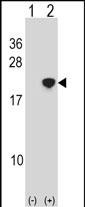
NANOS2 Antibody (Center)
Affinity Purified Rabbit Polyclonal Antibody (Pab)
- SPECIFICATION
- CITATIONS
- PROTOCOLS
- BACKGROUND

Application
| WB, E |
|---|---|
| Primary Accession | P60321 |
| Other Accession | NP_001025032.1 |
| Reactivity | Human |
| Host | Rabbit |
| Clonality | Polyclonal |
| Isotype | Rabbit IgG |
| Calculated MW | 15132 Da |
| Antigen Region | 58-85 aa |
| Gene ID | 339345 |
|---|---|
| Other Names | Nanos homolog 2, NOS-2, NANOS2, NOS2 |
| Target/Specificity | This NANOS2 antibody is generated from rabbits immunized with a KLH conjugated synthetic peptide between 58-85 amino acids from the Central region of human NANOS2. |
| Dilution | WB~~1:1000 E~~Use at an assay dependent concentration. |
| Format | Purified polyclonal antibody supplied in PBS with 0.09% (W/V) sodium azide. This antibody is purified through a protein A column, followed by peptide affinity purification. |
| Storage | Maintain refrigerated at 2-8°C for up to 2 weeks. For long term storage store at -20°C in small aliquots to prevent freeze-thaw cycles. |
| Precautions | NANOS2 Antibody (Center) is for research use only and not for use in diagnostic or therapeutic procedures. |
| Name | NANOS2 |
|---|---|
| Synonyms | NOS2 |
| Function | Plays a key role in the sexual differentiation of germ cells by promoting the male fate but suppressing the female fate. Represses the female fate pathways by suppressing meiosis, which in turn results in the promotion of the male fate. Maintains the suppression of meiosis by preventing STRA8 expression, which is required for premeiotic DNA replication, after CYP26B1 is decreased. Regulates the localization of the CCR4-NOT deadenylation complex to P-bodies and plays a role in recruiting the complex to trigger the degradation of mRNAs involved in meiosis. Required for the maintenance of the spermatogonial stem cell population. Not essential for the assembly of P-bodies but is required for the maintenance of their normal state (By similarity). |
| Cellular Location | Cytoplasm. Cytoplasm, P-body. Cytoplasm, perinuclear region. Note=Localizes at P-bodies during gonocyte development (By similarity). More abundant in perinuclear region of the cytoplasm of the germ cells of the adult testis |
| Tissue Location | Testis and ovary. Expression found in several spermatogenic stages: in cells on the periphery of the tubules which could correspond to spermatogonia, in spermatocytes and in round spermatids (at protein level). |

Thousands of laboratories across the world have published research that depended on the performance of antibodies from Abcepta to advance their research. Check out links to articles that cite our products in major peer-reviewed journals, organized by research category.
info@abcepta.com, and receive a free "I Love Antibodies" mug.

Provided below are standard protocols that you may find useful for product applications.
Background
NANOS2 is required to support proliferation and self-renewal of proximal germ cells in males only. Probably regulates translation of specific mRNAs by associating with the 3'-UTR of mRNA targets. Essential for spermatogonia formation (By similarity).
References
Kusz, K.M., et al. Mol. Hum. Reprod. 15(3):165-171(2009)
Chang, H.R., et al. Arch. Dermatol. Res. 295(7):293-296(2003)
Tsuda, M., et al. Science 301(5637):1239-1241(2003)
Jaruzelska, J., et al. Dev. Genes Evol. 213(3):120-126(2003)
If you have used an Abcepta product and would like to share how it has performed, please click on the "Submit Review" button and provide the requested information. Our staff will examine and post your review and contact you if needed.
If you have any additional inquiries please email technical services at tech@abcepta.com.
Ordering Information
Other Products
Shipping Information
