


 Foundational characteristics of cancer include proliferation, angiogenesis, migration, evasion of apoptosis, and cellular immortality. Find key markers for these cellular processes and antibodies to detect them.
 The SUMOplot™ Analysis Program predicts and scores sumoylation sites in your protein. SUMOylation is a post-translational modification involved in various cellular processes, such as nuclear-cytosolic transport, transcriptional regulation, apoptosis, protein stability, response to stress, and progression through the cell cycle.
 The Autophagy Receptor Motif Plotter predicts and scores autophagy receptor binding sites in your protein. Identifying proteins connected to this pathway is critical to understanding the role of autophagy in physiological as well as pathological processes such as development, differentiation, neurodegenerative diseases, stress, infection, and cancer.
TREML1 Antibody (C-term)
Affinity Purified Rabbit Polyclonal Antibody (Pab)
- SPECIFICATION
- CITATIONS
- PROTOCOLS
- BACKGROUND

Application
| WB, E |
|---|---|
| Primary Accession | Q86YW5 |
| Other Accession | NP_835468.1 |
| Reactivity | Human, Mouse |
| Host | Rabbit |
| Clonality | Polyclonal |
| Isotype | Rabbit IgG |
| Calculated MW | 32679 Da |
| Antigen Region | 215-241 aa |
| Gene ID | 340205 |
|---|---|
| Other Names | Trem-like transcript 1 protein, TLT-1, Triggering receptor expressed on myeloid cells-like protein 1, TREML1, TLT1 |
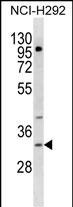
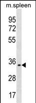
| Target/Specificity | This TREML1 antibody is generated from rabbits immunized with a KLH conjugated synthetic peptide between 215-241 amino acids from the C-terminal region of human TREML1. |
| Dilution | WB~~1:1000 E~~Use at an assay dependent concentration. |
| Format | Purified polyclonal antibody supplied in PBS with 0.09% (W/V) sodium azide. This antibody is purified through a protein A column, followed by peptide affinity purification. |
| Storage | Maintain refrigerated at 2-8°C for up to 2 weeks. For long term storage store at -20°C in small aliquots to prevent freeze-thaw cycles. |
| Precautions | TREML1 Antibody (C-term) is for research use only and not for use in diagnostic or therapeutic procedures. |
| Name | TREML1 |
|---|---|
| Synonyms | TLT1 |
| Function | Cell surface receptor that may play a role in the innate and adaptive immune response. |
| Cellular Location | Cell membrane; Single-pass type I membrane protein Cytoplasm. Note=Sequestered in cytoplasmic vesicles in resting platelets (PubMed:15100151) Transported to the cell surface after stimulation by thrombin (PubMed:15100151). Soluble fragments can be released into the serum by proteolysis (PubMed:16505478) |
| Tissue Location | Detected in platelets, monocytic leukemia and in T- cell leukemia. |

Thousands of laboratories across the world have published research that depended on the performance of antibodies from Abcepta to advance their research. Check out links to articles that cite our products in major peer-reviewed journals, organized by research category.
info@abcepta.com, and receive a free "I Love Antibodies" mug.

Provided below are standard protocols that you may find useful for product applications.
Background
TREML1 is located in a gene cluster on chromosome 6 with the single Ig variable (IgV) domain activating receptors TREM1 (MIM 605085) and TREM2 (MIM 605086), but it has distinct structural and functional properties. TREML1 enhances calcium signaling in an SHP2 (PTPN11; MIM 176876)-dependent manner (Allcock et al., 2003 [PubMed 12645956]; Barrow et al., 2004 [PubMed 15128762]).[supplied by OMIM].
References
Morales, J., et al. Blood Coagul. Fibrinolysis 21(3):229-236(2010)
Washington, A.V., et al. J. Clin. Invest. 119(6):1489-1501(2009)
Ford, J.W., et al. Curr. Opin. Immunol. 21(1):38-46(2009)
Nurden, A.T., et al. Thromb. Haemost. 100(1):45-51(2008)
Giomarelli, B., et al. Thromb. Haemost. 97(6):955-963(2007)
If you have used an Abcepta product and would like to share how it has performed, please click on the "Submit Review" button and provide the requested information. Our staff will examine and post your review and contact you if needed.
If you have any additional inquiries please email technical services at tech@abcepta.com.
Ordering Information
Other Products
Shipping Information
