


 Foundational characteristics of cancer include proliferation, angiogenesis, migration, evasion of apoptosis, and cellular immortality. Find key markers for these cellular processes and antibodies to detect them.
 The SUMOplot™ Analysis Program predicts and scores sumoylation sites in your protein. SUMOylation is a post-translational modification involved in various cellular processes, such as nuclear-cytosolic transport, transcriptional regulation, apoptosis, protein stability, response to stress, and progression through the cell cycle.
 The Autophagy Receptor Motif Plotter predicts and scores autophagy receptor binding sites in your protein. Identifying proteins connected to this pathway is critical to understanding the role of autophagy in physiological as well as pathological processes such as development, differentiation, neurodegenerative diseases, stress, infection, and cancer.
Rat Stra6 Antibody (C-term)
Affinity Purified Rabbit Polyclonal Antibody (Pab)
- SPECIFICATION
- CITATIONS
- PROTOCOLS
- BACKGROUND

Application
| WB, E |
|---|---|
| Primary Accession | Q4QR83 |
| Other Accession | NP_001025095.1 |
| Reactivity | Rat |
| Host | Rabbit |
| Clonality | Polyclonal |
| Isotype | Rabbit IgG |
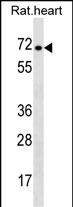
| Calculated MW | 73763 Da |
| Antigen Region | 483-509 aa |
| Gene ID | 363071 |
|---|---|
| Other Names | Stimulated by retinoic acid gene 6 protein homolog, Stra6 |
| Target/Specificity | This Rat Stra6 antibody is generated from rabbits immunized with a KLH conjugated synthetic peptide between 483-509 amino acids from the C-terminal region of rat Stra6. |
| Dilution | WB~~1:1000 E~~Use at an assay dependent concentration. |
| Format | Purified polyclonal antibody supplied in PBS with 0.09% (W/V) sodium azide. This antibody is purified through a protein A column, followed by peptide affinity purification. |
| Storage | Maintain refrigerated at 2-8°C for up to 2 weeks. For long term storage store at -20°C in small aliquots to prevent freeze-thaw cycles. |
| Precautions | Rat Stra6 Antibody (C-term) is for research use only and not for use in diagnostic or therapeutic procedures. |
| Name | Stra6 |
|---|---|
| Function | Functions as a retinol transporter. Accepts all-trans retinol from the extracellular retinol-binding protein RBP4, facilitates retinol transport across the cell membrane, and then transfers retinol to the cytoplasmic retinol-binding protein RBP1. Retinol uptake is enhanced by LRAT, an enzyme that converts retinol to all-trans retinyl esters, the storage forms of vitamin A. Contributes to the activation of a signaling cascade that depends on retinol transport and LRAT- dependent generation of retinol metabolites that then trigger activation of JAK2 and its target STAT5, and ultimately increase the expression of SOCS3 and inhibit cellular responses to insulin. Important for the homeostasis of vitamin A and its derivatives, such as retinoic acid. STRA6-mediated transport is particularly important in the eye, and under conditions of dietary vitamin A deficiency. Does not transport retinoic acid. |
| Cellular Location | Cell membrane {ECO:0000250|UniProtKB:Q0V8E7}; Multi-pass membrane protein {ECO:0000250|UniProtKB:Q0V8E7}. Note=In the retinal pigment epithelium localizes to the basolateral membrane {ECO:0000250|UniProtKB:Q0V8E7} |

Thousands of laboratories across the world have published research that depended on the performance of antibodies from Abcepta to advance their research. Check out links to articles that cite our products in major peer-reviewed journals, organized by research category.
info@abcepta.com, and receive a free "I Love Antibodies" mug.

Provided below are standard protocols that you may find useful for product applications.
Background
Stra6 may act as a high-affinity cell-surface receptor for the complex retinol-retinol binding protein (RBP/RBP4). Acts by removing retinol from RBP/RBP4 and transports it across the plasma membrane, where it can be metabolized. This mechanism does not depend on endocytosis. Binds to RBP/RBP4 with high affinity. Increases cellular retinol uptake from the retinol-RBP complex (By similarity).
References
Strausberg, R.L., et al. Proc. Natl. Acad. Sci. U.S.A. 99(26):16899-16903(2002)
If you have used an Abcepta product and would like to share how it has performed, please click on the "Submit Review" button and provide the requested information. Our staff will examine and post your review and contact you if needed.
If you have any additional inquiries please email technical services at tech@abcepta.com.
Ordering Information
Other Products
Shipping Information
