


 Foundational characteristics of cancer include proliferation, angiogenesis, migration, evasion of apoptosis, and cellular immortality. Find key markers for these cellular processes and antibodies to detect them.
 The SUMOplot™ Analysis Program predicts and scores sumoylation sites in your protein. SUMOylation is a post-translational modification involved in various cellular processes, such as nuclear-cytosolic transport, transcriptional regulation, apoptosis, protein stability, response to stress, and progression through the cell cycle.
 The Autophagy Receptor Motif Plotter predicts and scores autophagy receptor binding sites in your protein. Identifying proteins connected to this pathway is critical to understanding the role of autophagy in physiological as well as pathological processes such as development, differentiation, neurodegenerative diseases, stress, infection, and cancer.
KAZ Antibody (C-term)
Affinity Purified Rabbit Polyclonal Antibody (Pab)
- SPECIFICATION
- CITATIONS
- PROTOCOLS
- BACKGROUND

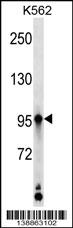
Application
| WB, E |
|---|---|
| Primary Accession | Q674X7 |
| Other Accession | NP_001017999.1 |
| Reactivity | Human |
| Host | Rabbit |
| Clonality | Polyclonal |
| Isotype | Rabbit IgG |
| Calculated MW | 86351 Da |
| Antigen Region | 711-739 aa |
| Gene ID | 23254 |
|---|---|
| Other Names | Kazrin, KAZN, KAZ, KIAA1026 |
| Target/Specificity | This KAZ antibody is generated from rabbits immunized with a KLH conjugated synthetic peptide between 711-739 amino acids from the C-terminal region of human KAZ. |
| Dilution | WB~~1:1000 E~~Use at an assay dependent concentration. |
| Format | Purified polyclonal antibody supplied in PBS with 0.09% (W/V) sodium azide. This antibody is purified through a protein A column, followed by peptide affinity purification. |
| Storage | Maintain refrigerated at 2-8°C for up to 2 weeks. For long term storage store at -20°C in small aliquots to prevent freeze-thaw cycles. |
| Precautions | KAZ Antibody (C-term) is for research use only and not for use in diagnostic or therapeutic procedures. |
| Name | KAZN (HGNC:29173) |
|---|---|
| Function | Component of the cornified envelope of keratinocytes. May be involved in the interplay between adherens junctions and desmosomes. The function in the nucleus is not known. |
| Cellular Location | Cytoplasm, cytoskeleton. [Isoform 3]: Cytoplasm. Cell junction, desmosome. Nucleus. Note=Observed at the apical plasma membrane of keratinocytes Partially colocalizes with PPL and DP at desmosomes, and with PP at the interdesmosomal plasma membrane. Colocalizes with cortical actin-based membrane structures |
| Tissue Location | Isoform 2, isoform 3 and isoform 4 are expressed in several cell lines including keratinocytes and bladder and epidermoid carcinoma (at protein level). Isoform 2, isoform 3 and isoform 4 are expressed in hair follicle and interfollicular epidermis (at protein level). |

Thousands of laboratories across the world have published research that depended on the performance of antibodies from Abcepta to advance their research. Check out links to articles that cite our products in major peer-reviewed journals, organized by research category.
info@abcepta.com, and receive a free "I Love Antibodies" mug.

Provided below are standard protocols that you may find useful for product applications.
Background
Component of the cornified envelope of keratinocytes. May be involved in the interplay between adherens junctions and desmosomes. The function in the nucleus is not known.
References
Rose, J.E., et al. Mol. Med. 16 (7-8), 247-253 (2010) :
Nachat, R., et al. J. Cell. Sci. 122 (PT 22), 4035-4041 (2009) :
Wang, Q., et al. Acta Biochim. Biophys. Sin. (Shanghai) 41(9):763-772(2009)
Sevilla, L.M., et al. J. Cell. Sci. 121 (PT 21), 3561-3569 (2008) :
Uhl, G.R., et al. Arch. Gen. Psychiatry 65(6):683-693(2008)
If you have used an Abcepta product and would like to share how it has performed, please click on the "Submit Review" button and provide the requested information. Our staff will examine and post your review and contact you if needed.
If you have any additional inquiries please email technical services at tech@abcepta.com.
Ordering Information
Other Products
Shipping Information
