


 Foundational characteristics of cancer include proliferation, angiogenesis, migration, evasion of apoptosis, and cellular immortality. Find key markers for these cellular processes and antibodies to detect them.
 The SUMOplot™ Analysis Program predicts and scores sumoylation sites in your protein. SUMOylation is a post-translational modification involved in various cellular processes, such as nuclear-cytosolic transport, transcriptional regulation, apoptosis, protein stability, response to stress, and progression through the cell cycle.
 The Autophagy Receptor Motif Plotter predicts and scores autophagy receptor binding sites in your protein. Identifying proteins connected to this pathway is critical to understanding the role of autophagy in physiological as well as pathological processes such as development, differentiation, neurodegenerative diseases, stress, infection, and cancer.
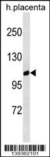
GPRC6A Antibody (Center)
Affinity Purified Rabbit Polyclonal Antibody (Pab)
- SPECIFICATION
- CITATIONS
- PROTOCOLS
- BACKGROUND

Application
| WB, E |
|---|---|
| Primary Accession | Q5T6X5 |
| Other Accession | NP_683766.2 |
| Reactivity | Human |
| Host | Rabbit |
| Clonality | Polyclonal |
| Isotype | Rabbit IgG |
| Calculated MW | 104753 Da |
| Antigen Region | 489-517 aa |
| Gene ID | 222545 |
|---|---|
| Other Names | G-protein coupled receptor family C group 6 member A, hGPRC6A, G-protein coupled receptor GPCR33, hGPCR33, GPRC6A |
| Target/Specificity | This GPRC6A antibody is generated from rabbits immunized with a KLH conjugated synthetic peptide between 489-517 amino acids from the Central region of human GPRC6A. |
| Dilution | WB~~1:1000 E~~Use at an assay dependent concentration. |
| Format | Purified polyclonal antibody supplied in PBS with 0.09% (W/V) sodium azide. This antibody is purified through a protein A column, followed by peptide affinity purification. |
| Storage | Maintain refrigerated at 2-8°C for up to 2 weeks. For long term storage store at -20°C in small aliquots to prevent freeze-thaw cycles. |
| Precautions | GPRC6A Antibody (Center) is for research use only and not for use in diagnostic or therapeutic procedures. |
| Name | GPRC6A |
|---|---|
| Function | Receptor activated by multiple ligands, including osteocalcin (BGLAP), basic amino acids, and various cations (PubMed:15576628). Activated by amino acids with a preference for basic amino acids such as L-Lys, L-Arg and L-ornithine but also by small and polar amino acids (PubMed:15576628). The L-alpha amino acids respond is augmented by divalent cations Ca(2+) and Mg(2+) (By similarity). Seems to act through a G(q)/G(11) and G(i)-coupled pathway (By similarity). Regulates testosterone production by acting as a ligand for uncarboxylated osteocalcin hormone: osteocalcin-binding at the surface of Leydig cells initiates a signaling response that promotes the expression of enzymes required for testosterone synthesis in a CREB- dependent manner (By similarity). Mediates the non-genomic effects of androgens in multiple tissue (By similarity). May coordinate nutritional and hormonal anabolic signals through the sensing of extracellular amino acids, osteocalcin, divalent ions and its responsiveness to anabolic steroids (PubMed:20947496). |
| Cellular Location | Cell membrane {ECO:0000250|UniProtKB:Q8K4Z6}; Multi-pass membrane protein {ECO:0000250|UniProtKB:Q8K4Z6} |
| Tissue Location | Isoform 1 is expressed at high level in brain, skeletal muscle, testis, bone, calvaria, osteoblasts and leukocytes Expressed at intermediate level in liver, heart, kidney and spleen Expressed at low level in lung, pancreas, placenta and ovary. Not detected in thymus, prostate, small intestine, tongue and colon Isoform 1 and isoform 2 are expressed in kidney at the same level Isoform 2 is expressed at lower level than isoform 1 in the other tissues. |

Thousands of laboratories across the world have published research that depended on the performance of antibodies from Abcepta to advance their research. Check out links to articles that cite our products in major peer-reviewed journals, organized by research category.
info@abcepta.com, and receive a free "I Love Antibodies" mug.

Provided below are standard protocols that you may find useful for product applications.
Background
Members of family C of the G protein-coupled receptor (GPCR) superfamily, such as GPRC6A, are characterized by an evolutionarily conserved amino acid-sensing motif linked to an intramembranous 7-transmembrane loop region. Several members of GPCR family C, including GPRC6A, also have a long N-terminal domain (summary by Pi et al., 2005 [PubMed 16199532]).
References
Giroux, S., et al. Bone 47(5):975-981(2010)
Takata, R., et al. Nat. Genet. 42(9):751-754(2010)
Pi, M., et al. J. Bone Miner. Res. 25(5):1092-1102(2010)
Dowal, L., et al. J. Biol. Chem. 281(33):23999-24014(2006)
Pi, M., et al. J. Biol. Chem. 280(48):40201-40209(2005)
If you have used an Abcepta product and would like to share how it has performed, please click on the "Submit Review" button and provide the requested information. Our staff will examine and post your review and contact you if needed.
If you have any additional inquiries please email technical services at tech@abcepta.com.
Ordering Information
Other Products
Shipping Information
