


 Foundational characteristics of cancer include proliferation, angiogenesis, migration, evasion of apoptosis, and cellular immortality. Find key markers for these cellular processes and antibodies to detect them.
 The SUMOplot™ Analysis Program predicts and scores sumoylation sites in your protein. SUMOylation is a post-translational modification involved in various cellular processes, such as nuclear-cytosolic transport, transcriptional regulation, apoptosis, protein stability, response to stress, and progression through the cell cycle.
 The Autophagy Receptor Motif Plotter predicts and scores autophagy receptor binding sites in your protein. Identifying proteins connected to this pathway is critical to understanding the role of autophagy in physiological as well as pathological processes such as development, differentiation, neurodegenerative diseases, stress, infection, and cancer.
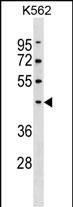
PELI1 Antibody (Center)
Affinity Purified Rabbit Polyclonal Antibody (Pab)
- SPECIFICATION
- CITATIONS
- PROTOCOLS
- BACKGROUND

Application
| WB, E |
|---|---|
| Primary Accession | Q96FA3 |
| Other Accession | Q8C669, NP_065702.2 |
| Reactivity | Human |
| Predicted | Mouse |
| Host | Rabbit |
| Clonality | Polyclonal |
| Isotype | Rabbit IgG |
| Calculated MW | 46286 Da |
| Antigen Region | 177-204 aa |
| Gene ID | 57162 |
|---|---|
| Other Names | E3 ubiquitin-protein ligase pellino homolog 1, Pellino-1, 632-, Pellino-related intracellular-signaling molecule, PELI1, PRISM |
| Target/Specificity | This PELI1 antibody is generated from rabbits immunized with a KLH conjugated synthetic peptide between 177-204 amino acids from the Central region of human PELI1. |
| Dilution | WB~~1:1000 E~~Use at an assay dependent concentration. |
| Format | Purified polyclonal antibody supplied in PBS with 0.09% (W/V) sodium azide. This antibody is purified through a protein A column, followed by peptide affinity purification. |
| Storage | Maintain refrigerated at 2-8°C for up to 2 weeks. For long term storage store at -20°C in small aliquots to prevent freeze-thaw cycles. |
| Precautions | PELI1 Antibody (Center) is for research use only and not for use in diagnostic or therapeutic procedures. |
| Name | PELI1 {ECO:0000303|PubMed:30952868} |
|---|---|
| Synonyms | PRISM |
| Function | E3 ubiquitin ligase catalyzing the covalent attachment of ubiquitin moieties onto substrate proteins (PubMed:12496252, PubMed:17675297, PubMed:29883609, PubMed:30952868). Involved in the TLR and IL-1 signaling pathways via interaction with the complex containing IRAK kinases and TRAF6 (PubMed:12496252, PubMed:17675297). Acts as a positive regulator of inflammatory response in microglia through activation of NF-kappa-B and MAP kinase (By similarity). Mediates 'Lys- 63'-linked polyubiquitination of IRAK1 allowing subsequent NF-kappa-B activation (PubMed:12496252, PubMed:17675297). Conjugates 'Lys-63'- linked ubiquitin chains to the adapter protein ASC/PYCARD, which in turn is crucial for NLRP3 inflammasome activation (PubMed:34706239). Mediates 'Lys-48'-linked polyubiquitination of RIPK3 leading to its subsequent proteasome-dependent degradation; preferentially recognizes and mediates the degradation of the 'Thr-182' phosphorylated form of RIPK3 (PubMed:29883609). Negatively regulates necroptosis by reducing RIPK3 expression (PubMed:29883609). Mediates 'Lys-63'-linked ubiquitination of RIPK1 (PubMed:29883609). Following phosphorylation by ATM, catalyzes 'Lys-63'-linked ubiquitination of NBN, promoting DNA repair via homologous recombination (PubMed:30952868). Negatively regulates activation of the metabolic mTORC1 signaling pathway by mediating 'Lys-63'-linked ubiquitination of mTORC1-inhibitory protein TSC1 and thereby promoting TSC1/TSC2 complex stability (PubMed:33215753). |
| Cellular Location | Chromosome. Note=Localizes to DNA double-strand breaks (DSBs) in response to DNA damage. |
| Tissue Location | Expressed at high levels in normal skin but decreased in keratinocytes from toxic epidermal necrolysis (TEN) patients (at protein level). |

Thousands of laboratories across the world have published research that depended on the performance of antibodies from Abcepta to advance their research. Check out links to articles that cite our products in major peer-reviewed journals, organized by research category.
info@abcepta.com, and receive a free "I Love Antibodies" mug.

Provided below are standard protocols that you may find useful for product applications.
Background
Scaffold protein involved in the IL-1 signaling pathway via its interaction with the complex containing IRAK kinases and TRAF6. Required for NF-kappa-B activation and IL-8 gene expression in response to IL-1.
References
Ordureau, A., et al. Biochem. J. 409(1):43-52(2008)
Butler, M.P., et al. J. Biol. Chem. 282(41):29729-29737(2007)
Lamesch, P., et al. Genomics 89(3):307-315(2007)
Choi, K.C., et al. Nat. Immunol. 7(10):1057-1065(2006)
Butler, M.P., et al. J. Biol. Chem. 280(30):27759-27768(2005)
If you have used an Abcepta product and would like to share how it has performed, please click on the "Submit Review" button and provide the requested information. Our staff will examine and post your review and contact you if needed.
If you have any additional inquiries please email technical services at tech@abcepta.com.
Ordering Information
Other Products
Shipping Information
