


 Foundational characteristics of cancer include proliferation, angiogenesis, migration, evasion of apoptosis, and cellular immortality. Find key markers for these cellular processes and antibodies to detect them.
 The SUMOplot™ Analysis Program predicts and scores sumoylation sites in your protein. SUMOylation is a post-translational modification involved in various cellular processes, such as nuclear-cytosolic transport, transcriptional regulation, apoptosis, protein stability, response to stress, and progression through the cell cycle.
 The Autophagy Receptor Motif Plotter predicts and scores autophagy receptor binding sites in your protein. Identifying proteins connected to this pathway is critical to understanding the role of autophagy in physiological as well as pathological processes such as development, differentiation, neurodegenerative diseases, stress, infection, and cancer.
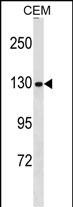
NCAPG Antibody (N-term)
Affinity Purified Rabbit Polyclonal Antibody (Pab)
- SPECIFICATION
- CITATIONS: 1
- PROTOCOLS
- BACKGROUND

Application
| WB, E |
|---|---|
| Primary Accession | Q9BPX3 |
| Other Accession | NP_071741.2 |
| Reactivity | Human |
| Host | Rabbit |
| Clonality | Polyclonal |
| Isotype | Rabbit IgG |
| Calculated MW | 114334 Da |
| Antigen Region | 117-145 aa |
| Gene ID | 64151 |
|---|---|
| Other Names | Condensin complex subunit 3, Chromosome-associated protein G, Condensin subunit CAP-G, hCAP-G, Melanoma antigen NY-MEL-3, Non-SMC condensin I complex subunit G, XCAP-G homolog, NCAPG, CAPG, NYMEL3 |
| Target/Specificity | This NCAPG antibody is generated from rabbits immunized with a KLH conjugated synthetic peptide between 117-145 amino acids from the N-terminal region of human NCAPG. |
| Dilution | WB~~1:1000 E~~Use at an assay dependent concentration. |
| Format | Purified polyclonal antibody supplied in PBS with 0.09% (W/V) sodium azide. This antibody is purified through a protein A column, followed by peptide affinity purification. |
| Storage | Maintain refrigerated at 2-8°C for up to 2 weeks. For long term storage store at -20°C in small aliquots to prevent freeze-thaw cycles. |
| Precautions | NCAPG Antibody (N-term) is for research use only and not for use in diagnostic or therapeutic procedures. |
| Name | NCAPG |
|---|---|
| Synonyms | CAPG, NYMEL3 |
| Function | Regulatory subunit of the condensin complex, a complex required for conversion of interphase chromatin into mitotic-like condense chromosomes. The condensin complex probably introduces positive supercoils into relaxed DNA in the presence of type I topoisomerases and converts nicked DNA into positive knotted forms in the presence of type II topoisomerases. |
| Cellular Location | Nucleus. Cytoplasm. Chromosome. Note=In interphase cells, the majority of the condensin complex is found in the cytoplasm, while a minority of the complex is associated with chromatin. A subpopulation of the complex however remains associated with chromosome foci in interphase cells. During mitosis, most of the condensin complex is associated with the chromatin. At the onset of prophase, the regulatory subunits of the complex are phosphorylated by CDK1, leading to condensin's association with chromosome arms and to chromosome condensation. Dissociation from chromosomes is observed in late telophase |
| Tissue Location | Highly expressed in testis. |

Provided below are standard protocols that you may find useful for product applications.
Background
Regulatory subunit of the condensin complex, a complex required for conversion of interphase chromatin into mitotic-like condense chromosomes. The condensin complex probably introduces positive supercoils into relaxed DNA in the presence of type I topoisomerases and converts nicked DNA into positive knotted forms in the presence of type II topoisomerases.
References
Okada, Y., et al. Hum. Mol. Genet. 19(11):2303-2312(2010)
Zhao, J., et al. BMC Med. Genet. 11, 96 (2010) :
Murphy, L.A., et al. Biochem. Biophys. Res. Commun. 377(3):1007-1011(2008)
Gudbjartsson, D.F., et al. Nat. Genet. 40(5):609-615(2008)
Sugiyama, N., et al. Mol. Cell Proteomics 6(6):1103-1109(2007)
If you have used an Abcepta product and would like to share how it has performed, please click on the "Submit Review" button and provide the requested information. Our staff will examine and post your review and contact you if needed.
If you have any additional inquiries please email technical services at tech@abcepta.com.

Ordering Information
Shipping Information
