


 Foundational characteristics of cancer include proliferation, angiogenesis, migration, evasion of apoptosis, and cellular immortality. Find key markers for these cellular processes and antibodies to detect them.
 The SUMOplot™ Analysis Program predicts and scores sumoylation sites in your protein. SUMOylation is a post-translational modification involved in various cellular processes, such as nuclear-cytosolic transport, transcriptional regulation, apoptosis, protein stability, response to stress, and progression through the cell cycle.
 The Autophagy Receptor Motif Plotter predicts and scores autophagy receptor binding sites in your protein. Identifying proteins connected to this pathway is critical to understanding the role of autophagy in physiological as well as pathological processes such as development, differentiation, neurodegenerative diseases, stress, infection, and cancer.
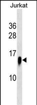
ATP6V1F Antibody (C-term)
Affinity Purified Rabbit Polyclonal Antibody (Pab)
- SPECIFICATION
- CITATIONS
- PROTOCOLS
- BACKGROUND

Application
| WB, E |
|---|---|
| Primary Accession | Q16864 |
| Other Accession | Q28029, NP_004222.2 |
| Reactivity | Human |
| Predicted | Bovine |
| Host | Rabbit |
| Clonality | Polyclonal |
| Isotype | Rabbit IgG |
| Calculated MW | 13370 Da |
| Antigen Region | 82-111 aa |
| Gene ID | 9296 |
|---|---|
| Other Names | V-type proton ATPase subunit F, V-ATPase subunit F, V-ATPase 14 kDa subunit, Vacuolar proton pump subunit F, ATP6V1F, ATP6S14, VATF |
| Target/Specificity | This ATP6V1F antibody is generated from rabbits immunized with a KLH conjugated synthetic peptide between 82-111 amino acids from the C-terminal region of human ATP6V1F. |
| Dilution | WB~~1:1000 E~~Use at an assay dependent concentration. |
| Format | Purified polyclonal antibody supplied in PBS with 0.09% (W/V) sodium azide. This antibody is purified through a protein A column, followed by peptide affinity purification. |
| Storage | Maintain refrigerated at 2-8°C for up to 2 weeks. For long term storage store at -20°C in small aliquots to prevent freeze-thaw cycles. |
| Precautions | ATP6V1F Antibody (C-term) is for research use only and not for use in diagnostic or therapeutic procedures. |
| Name | ATP6V1F |
|---|---|
| Synonyms | ATP6S14, VATF |
| Function | Subunit of the V1 complex of vacuolar(H+)-ATPase (V-ATPase), a multisubunit enzyme composed of a peripheral complex (V1) that hydrolyzes ATP and a membrane integral complex (V0) that translocates protons (PubMed:33065002). V-ATPase is responsible for acidifying and maintaining the pH of intracellular compartments and in some cell types, is targeted to the plasma membrane, where it is responsible for acidifying the extracellular environment (By similarity). |
| Cellular Location | Cytoplasmic vesicle, secretory vesicle, synaptic vesicle membrane {ECO:0000250|UniProtKB:P50408}; Peripheral membrane protein. Cytoplasmic vesicle, clathrin-coated vesicle membrane {ECO:0000250|UniProtKB:P50408}; Peripheral membrane protein |

Thousands of laboratories across the world have published research that depended on the performance of antibodies from Abcepta to advance their research. Check out links to articles that cite our products in major peer-reviewed journals, organized by research category.
info@abcepta.com, and receive a free "I Love Antibodies" mug.

Provided below are standard protocols that you may find useful for product applications.
Background
This gene encodes a component of vacuolar ATPase (V-ATPase), a multisubunit enzyme that mediates acidification of eukaryotic intracellular organelles. V-ATPase dependent organelle acidification is necessary for such intracellular processes as protein sorting, zymogen activation, receptor-mediated endocytosis, and synaptic vesicle proton gradient generation. V-ATPase is composed of a cytosolic V1 domain and a transmembrane V0 domain. The V1 domain consists of three A and three B subunits, two G subunits plus the C, D, E, F, and H subunits. The V1 domain contains the ATP catalytic site. The V0 domain consists of five different subunits: a, c, c', c', and d. Additional isoforms of many of the V1 and V0 subunit proteins are encoded by multiple genes or alternatively spliced transcript variants. This encoded protein is the V1 domain F subunit protein.
References
Supino, R., et al. Ann. N. Y. Acad. Sci. 1171, 606-616 (2009) :
Smith, A.N., et al. J. Bioenerg. Biomembr. 40(4):371-380(2008)
Morel, N. Biol. Cell 95(7):453-457(2003)
Smith, A.N., et al. Mol. Cell 12(4):801-803(2003)
Kawasaki-Nishi, S., et al. FEBS Lett. 545(1):76-85(2003)
If you have used an Abcepta product and would like to share how it has performed, please click on the "Submit Review" button and provide the requested information. Our staff will examine and post your review and contact you if needed.
If you have any additional inquiries please email technical services at tech@abcepta.com.
Ordering Information
Other Products
Shipping Information
