


 Foundational characteristics of cancer include proliferation, angiogenesis, migration, evasion of apoptosis, and cellular immortality. Find key markers for these cellular processes and antibodies to detect them.
 The SUMOplot™ Analysis Program predicts and scores sumoylation sites in your protein. SUMOylation is a post-translational modification involved in various cellular processes, such as nuclear-cytosolic transport, transcriptional regulation, apoptosis, protein stability, response to stress, and progression through the cell cycle.
 The Autophagy Receptor Motif Plotter predicts and scores autophagy receptor binding sites in your protein. Identifying proteins connected to this pathway is critical to understanding the role of autophagy in physiological as well as pathological processes such as development, differentiation, neurodegenerative diseases, stress, infection, and cancer.
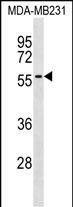
AKAP5 Antibody (Center)
Affinity Purified Rabbit Polyclonal Antibody (Pab)
- SPECIFICATION
- CITATIONS
- PROTOCOLS
- BACKGROUND

Application
| WB, E |
|---|---|
| Primary Accession | P24588 |
| Other Accession | NP_004848.3 |
| Reactivity | Human |
| Host | Rabbit |
| Clonality | Polyclonal |
| Isotype | Rabbit IgG |
| Calculated MW | 47088 Da |
| Antigen Region | 115-143 aa |
| Gene ID | 9495 |
|---|---|
| Other Names | A-kinase anchor protein 5, AKAP-5, A-kinase anchor protein 79 kDa, AKAP 79, H21, cAMP-dependent protein kinase regulatory subunit II high affinity-binding protein, AKAP5, AKAP79 |
| Target/Specificity | This AKAP5 antibody is generated from rabbits immunized with a KLH conjugated synthetic peptide between 115-143 amino acids from the Central region of human AKAP5. |
| Dilution | WB~~1:1000 E~~Use at an assay dependent concentration. |
| Format | Purified polyclonal antibody supplied in PBS with 0.09% (W/V) sodium azide. This antibody is purified through a protein A column, followed by peptide affinity purification. |
| Storage | Maintain refrigerated at 2-8°C for up to 2 weeks. For long term storage store at -20°C in small aliquots to prevent freeze-thaw cycles. |
| Precautions | AKAP5 Antibody (Center) is for research use only and not for use in diagnostic or therapeutic procedures. |
| Name | AKAP5 |
|---|---|
| Synonyms | AKAP79 |
| Function | Multivalent scaffold protein that anchors the cAMP-dependent protein kinase/PKA to cytoskeletal and/or organelle-associated proteins, targeting the signal carried by cAMP to specific intracellular effectors (PubMed:1512224). Association with the beta2- adrenergic receptor (beta2-AR) not only regulates beta2-AR signaling pathway, but also the activation by PKA by switching off the beta2-AR signaling cascade. Plays a role in long term synaptic potentiation by regulating protein trafficking from the dendritic recycling endosomes to the plasma membrane and controlling both structural and functional plasticity at excitatory synapses (PubMed:25589740). In hippocampal pyramidal neurons, recruits KCNK2/TREK-1 channel at postsynaptic dense bodies microdomains and converts it to a leak channel no longer sensitive to stimulation by arachidonic acid, acidic pH or mechanical stress, nor inhibited by Gq-coupled receptors but still under the negative control of Gs-coupled receptors (By similarity). Associates with ORAI1 pore-forming subunit of CRAC channels in Ca(2+) signaling microdomains where it recruits NFATC2/NFAT1 and couples store-operated Ca(2+) influx to calmodulin and calcineurin signaling and activation of NFAT-dependent transcriptional responses (PubMed:33941685). |
| Cellular Location | Postsynaptic recycling endosome membrane; Lipid- anchor. Cell projection, dendrite {ECO:0000250|UniProtKB:D3YVF0}. Postsynaptic cell membrane {ECO:0000250|UniProtKB:D3YVF0}; Lipid-anchor. Note=Associates with lipid rafts. |
| Tissue Location | Predominantly in the cerebral cortex and the postsynaptic densities of the forebrain, and to a lesser extent in adrenal medulla, lung and anterior pituitary |

Thousands of laboratories across the world have published research that depended on the performance of antibodies from Abcepta to advance their research. Check out links to articles that cite our products in major peer-reviewed journals, organized by research category.
info@abcepta.com, and receive a free "I Love Antibodies" mug.

Provided below are standard protocols that you may find useful for product applications.
Background
The A-kinase anchor proteins (AKAPs) are a group of structurally diverse proteins, which have the common function of binding to the regulatory subunit of protein kinase A (PKA) and confining the holoenzyme to discrete locations within the cell. This gene encodes a member of the AKAP family. The encoded protein binds to the RII-beta regulatory subunit of PKA, and also to protein kinase C and the phosphatase calcineurin. It is predominantly expressed in cerebral cortex and may anchor the PKA protein at postsynaptic densities (PSD) and be involved in the regulation of postsynaptic events. It is also expressed in T lymphocytes and may function to inhibit interleukin-2 transcription by disrupting calcineurin-dependent dephosphorylation of NFAT.
References
Willoughby, D., et al. J. Biol. Chem. 285(26):20328-20342(2010)
Chen, M.H., et al. Cell. Signal. 21(1):136-142(2009)
Tavalin, S.J. J. Biol. Chem. 283(17):11445-11452(2008)
Correia, S.S., et al. Nat. Neurosci. 11(4):457-466(2008)
Chai, S., et al. J. Biol. Chem. 282(31):22668-22677(2007)
If you have used an Abcepta product and would like to share how it has performed, please click on the "Submit Review" button and provide the requested information. Our staff will examine and post your review and contact you if needed.
If you have any additional inquiries please email technical services at tech@abcepta.com.
Ordering Information
Other Products
Shipping Information
