


 Foundational characteristics of cancer include proliferation, angiogenesis, migration, evasion of apoptosis, and cellular immortality. Find key markers for these cellular processes and antibodies to detect them.
 The SUMOplot™ Analysis Program predicts and scores sumoylation sites in your protein. SUMOylation is a post-translational modification involved in various cellular processes, such as nuclear-cytosolic transport, transcriptional regulation, apoptosis, protein stability, response to stress, and progression through the cell cycle.
 The Autophagy Receptor Motif Plotter predicts and scores autophagy receptor binding sites in your protein. Identifying proteins connected to this pathway is critical to understanding the role of autophagy in physiological as well as pathological processes such as development, differentiation, neurodegenerative diseases, stress, infection, and cancer.
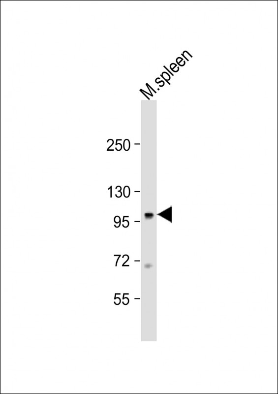
Mouse Nod1 Antibody (Center)
Affinity Purified Rabbit Polyclonal Antibody (Pab)
- SPECIFICATION
- CITATIONS
- PROTOCOLS
- BACKGROUND

Application
| WB, E |
|---|---|
| Primary Accession | Q8BHB0 |
| Other Accession | NP_001164478.1 |
| Reactivity | Mouse |
| Host | Rabbit |
| Clonality | Polyclonal |
| Isotype | Rabbit IgG |
| Calculated MW | 107740 Da |
| Antigen Region | 556-584 aa |
| Gene ID | 107607 |
|---|---|
| Other Names | Nucleotide-binding oligomerization domain-containing protein 1, Caspase recruitment domain-containing protein 4, Nod1, Card4 |
| Target/Specificity | This Mouse Nod1 antibody is generated from rabbits immunized with a KLH conjugated synthetic peptide between 556-584 amino acids from the Central region of mouse Nod1. |
| Dilution | WB~~1:1000 E~~Use at an assay dependent concentration. |
| Format | Purified polyclonal antibody supplied in PBS with 0.09% (W/V) sodium azide. This antibody is purified through a protein A column, followed by peptide affinity purification. |
| Storage | Maintain refrigerated at 2-8°C for up to 2 weeks. For long term storage store at -20°C in small aliquots to prevent freeze-thaw cycles. |
| Precautions | Mouse Nod1 Antibody (Center) is for research use only and not for use in diagnostic or therapeutic procedures. |
| Name | Nod1 {ECO:0000303|PubMed:16211083, ECO:0000312|MGI:MGI:1341839} |
|---|---|
| Function | Pattern recognition receptor (PRR) that detects bacterial peptidoglycan fragments and other danger signals and thus participates in both innate and adaptive immune responses (PubMed:12796777, PubMed:21715553). Specifically recognizes and binds gamma-D-glutamyl- meso-diaminopimelic acid (iE-DAP), a dipeptide present in peptidoglycan of Gram-negative bacteria (PubMed:12796777, PubMed:16211083). Preferentially binds iE-DAP in tetrapeptide-containing muropeptides (MurNAc-TetraDAP or TetraDAP) (PubMed:16211083). Ligand binding triggers oligomerization that facilitates the binding and subsequent activation of the proximal adapter receptor-interacting RIPK2 (By similarity). Following recruitment, RIPK2 undergoes 'Met-1'- (linear) and 'Lys-63'-linked polyubiquitination by E3 ubiquitin-protein ligases XIAP, BIRC2, BIRC3 and the LUBAC complex, becoming a scaffolding protein for downstream effectors, triggering activation of the NF- kappa-B and MAP kinases signaling (By similarity). This in turn leads to the transcriptional activation of hundreds of genes involved in immune response (By similarity). Also acts as a regulator of antiviral response elicited by dsRNA and the expression of RLR pathway members by targeting IFIH1 and TRAF3 to modulate the formation of IFIH1-MAVS and TRAF3-MAVS complexes leading to increased transcription of type I IFNs (By similarity). Also acts as a regulator of autophagy via its interaction with ATG16L1, possibly by recruiting ATG16L1 at the site of bacterial entry (PubMed:19898471). Besides recognizing pathogens, also involved in the endoplasmic reticulum stress response: acts by sensing and binding to the cytosolic metabolite sphingosine-1-phosphate generated in response to endoplasmic reticulum stress, initiating an inflammation process that leads to activation of the NF-kappa-B and MAP kinases signaling (PubMed:27007849). In addition, plays a role in insulin trafficking in beta cells in a cell-autonomous manner (PubMed:21715553, PubMed:31201384). Mechanistically, upon recognizing cognate ligands, NOD1 and RIPK2 localize to insulin vesicles where they recruit RAB1A to direct insulin trafficking through the cytoplasm (PubMed:31201384). |
| Cellular Location | Cell membrane {ECO:0000250|UniProtKB:Q9Y239}; Lipid-anchor {ECO:0000250|UniProtKB:Q9Y239}. Apical cell membrane {ECO:0000250|UniProtKB:Q9Y239}. Basolateral cell membrane {ECO:0000250|UniProtKB:Q9Y239}. Cytoplasm {ECO:0000250|UniProtKB:Q9Y239}. Note=Detected in the cytoplasm and at the cell membrane. Following bacterial infection, localizes to bacterial entry sites in the cell membrane. Recruited to the basolateral and apical membranes in polarized epithelial cells {ECO:0000250|UniProtKB:Q9Y239} |
| Tissue Location | Although ubiquitously expressed, NOD1 levels are more abundant in immune cells, the gastrointestinal tract, and adipose tissue. |

Thousands of laboratories across the world have published research that depended on the performance of antibodies from Abcepta to advance their research. Check out links to articles that cite our products in major peer-reviewed journals, organized by research category.
info@abcepta.com, and receive a free "I Love Antibodies" mug.

Provided below are standard protocols that you may find useful for product applications.
Background
Enhances caspase-9-mediated apoptosis. Induces NF-kappa-B activity via RIPK2 and IKK-gamma. Confers responsiveness to intracellular bacterial lipopolysaccharides (LPS). Forms an intracellular sensing system along with ARHGEF2 for the detection of microbial effectors during cell invasion by pathogens (By similarity).
References
Moreno, L., et al. Br. J. Pharmacol. 160(8):1997-2007(2010)
Iyer, J.K., et al. Infect. Immun. 78(6):2418-2428(2010)
Watanabe, T., et al. J. Clin. Invest. 120(5):1645-1662(2010)
Toma, C., et al. J. Immunol. 184(9):5287-5297(2010)
Shigeoka, A.A., et al. J. Immunol. 184(5):2297-2304(2010)
If you have used an Abcepta product and would like to share how it has performed, please click on the "Submit Review" button and provide the requested information. Our staff will examine and post your review and contact you if needed.
If you have any additional inquiries please email technical services at tech@abcepta.com.
Ordering Information
Other Products
Shipping Information
