


 Foundational characteristics of cancer include proliferation, angiogenesis, migration, evasion of apoptosis, and cellular immortality. Find key markers for these cellular processes and antibodies to detect them.
 The SUMOplot™ Analysis Program predicts and scores sumoylation sites in your protein. SUMOylation is a post-translational modification involved in various cellular processes, such as nuclear-cytosolic transport, transcriptional regulation, apoptosis, protein stability, response to stress, and progression through the cell cycle.
 The Autophagy Receptor Motif Plotter predicts and scores autophagy receptor binding sites in your protein. Identifying proteins connected to this pathway is critical to understanding the role of autophagy in physiological as well as pathological processes such as development, differentiation, neurodegenerative diseases, stress, infection, and cancer.
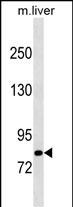
Mouse Cblb Antibody (C-term)
Affinity Purified Rabbit Polyclonal Antibody (Pab)
- SPECIFICATION
- CITATIONS
- PROTOCOLS
- BACKGROUND

Application
| WB, E |
|---|---|
| Primary Accession | Q3TTA7 |
| Other Accession | NP_001028410.1 |
| Reactivity | Mouse |
| Host | Rabbit |
| Clonality | Polyclonal |
| Isotype | Rabbit IgG |
| Calculated MW | 109092 Da |
| Antigen Region | 862-889 aa |
| Gene ID | 208650 |
|---|---|
| Other Names | E3 ubiquitin-protein ligase CBL-B, 632-, Casitas B-lineage lymphoma proto-oncogene b, SH3-binding protein CBL-B, Signal transduction protein CBL-B, Cblb |
| Target/Specificity | This Mouse Cblb antibody is generated from rabbits immunized with a KLH conjugated synthetic peptide between 862-889 amino acids from the C-terminal region of mouse Cblb. |
| Dilution | WB~~1:1000 E~~Use at an assay dependent concentration. |
| Format | Purified polyclonal antibody supplied in PBS with 0.09% (W/V) sodium azide. This antibody is purified through a protein A column, followed by peptide affinity purification. |
| Storage | Maintain refrigerated at 2-8°C for up to 2 weeks. For long term storage store at -20°C in small aliquots to prevent freeze-thaw cycles. |
| Precautions | Mouse Cblb Antibody (C-term) is for research use only and not for use in diagnostic or therapeutic procedures. |
| Name | Cblb |
|---|---|
| Function | E3 ubiquitin-protein ligase which accepts ubiquitin from specific E2 ubiquitin-conjugating enzymes, and transfers it to substrates, generally promoting their degradation by the proteasome. Negatively regulates TCR (T-cell receptor), BCR (B-cell receptor) and FCER1 (high affinity immunoglobulin epsilon receptor) signal transduction pathways. In naive T-cells, inhibits VAV1 activation upon TCR engagement and imposes a requirement for CD28 costimulation for proliferation and IL-2 production. Also acts by promoting PIK3R1/p85 ubiquitination, which impairs its recruitment to the TCR and subsequent activation. In activated T-cells, inhibits PLCG1 activation and calcium mobilization upon restimulation and promotes anergy. In B-cells, acts by ubiquitinating SYK and promoting its proteasomal degradation. Slightly promotes SRC ubiquitination. May be involved in EGFR ubiquitination and internalization. May be functionally coupled with the E2 ubiquitin-protein ligase UB2D3. In association with CBL, required for proper feedback inhibition of ciliary platelet-derived growth factor receptor-alpha (PDGFRA) signaling pathway via ubiquitination and internalization of PDGFRA (PubMed:29237719). |
| Cellular Location | Cytoplasm. Note=In adipocytes, translocates to the plasma membrane upon insulin stimulation |

Thousands of laboratories across the world have published research that depended on the performance of antibodies from Abcepta to advance their research. Check out links to articles that cite our products in major peer-reviewed journals, organized by research category.
info@abcepta.com, and receive a free "I Love Antibodies" mug.

Provided below are standard protocols that you may find useful for product applications.
Background
E3 ubiquitin-protein ligase which accepts ubiquitin from specific E2 ubiquitin-conjugating enzymes, and transfers it to substrates, generally promoting their degradation by the proteasome. Negatively regulates TCR (T-cell receptor), BCR (B-cell receptor) and FCER1 (high affinity immunoglobulin epsilon receptor) signal transduction pathways. In naive T-cells, inhibits VAV1 activation upon TCR engagement and imposes a requirement for CD28 costimulation for proliferation and IL-2 production. Also acts by promoting PIK3R1/p85 ubiquitination, which impairs its recruitment to the TCR and subsequent activation. In activated T-cells, inhibits PLCG1 activation and calcium mobilization upon restimulation and promotes anergy. In B-cells, acts by ubiquitinating SYK and promoting its proteasomal degradation. May also be involved in EGFR ubiquitination and internalization.
References
Wilson, B.G., et al. Cancer Cell 18(4):316-328(2010)
Stromnes, I.M., et al. J. Clin. Invest. 120(10):3722-3734(2010)
Naramura, M., et al. Proc. Natl. Acad. Sci. U.S.A. 107(37):16274-16279(2010)
Teh, C.E., et al. Proc. Natl. Acad. Sci. U.S.A. 107(33):14709-14714(2010)
Huang, H., et al. Immunity 33(1):60-70(2010)
If you have used an Abcepta product and would like to share how it has performed, please click on the "Submit Review" button and provide the requested information. Our staff will examine and post your review and contact you if needed.
If you have any additional inquiries please email technical services at tech@abcepta.com.
Ordering Information
Other Products
Shipping Information
