


 Foundational characteristics of cancer include proliferation, angiogenesis, migration, evasion of apoptosis, and cellular immortality. Find key markers for these cellular processes and antibodies to detect them.
 The SUMOplot™ Analysis Program predicts and scores sumoylation sites in your protein. SUMOylation is a post-translational modification involved in various cellular processes, such as nuclear-cytosolic transport, transcriptional regulation, apoptosis, protein stability, response to stress, and progression through the cell cycle.
 The Autophagy Receptor Motif Plotter predicts and scores autophagy receptor binding sites in your protein. Identifying proteins connected to this pathway is critical to understanding the role of autophagy in physiological as well as pathological processes such as development, differentiation, neurodegenerative diseases, stress, infection, and cancer.
APC Antibody (Center)
Affinity Purified Rabbit Polyclonal Antibody (Pab)
- SPECIFICATION
- CITATIONS
- PROTOCOLS
- BACKGROUND

Application
| WB, E |
|---|---|
| Primary Accession | P25054 |
| Other Accession | NP_000029.2 |
| Reactivity | Mouse |
| Host | Rabbit |
| Clonality | Polyclonal |
| Isotype | Rabbit IgG |
| Calculated MW | 311646 Da |
| Antigen Region | 1148-1176 aa |
| Gene ID | 324 |
|---|---|
| Other Names | Adenomatous polyposis coli protein, Protein APC, Deleted in polyposis 25, APC, DP25 |
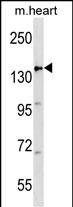
| Target/Specificity | This APC antibody is generated from rabbits immunized with a KLH conjugated synthetic peptide between 1148-1176 amino acids from the Central region of human APC. |
| Dilution | WB~~1:1000 E~~Use at an assay dependent concentration. |
| Format | Purified polyclonal antibody supplied in PBS with 0.09% (W/V) sodium azide. This antibody is purified through a protein A column, followed by peptide affinity purification. |
| Storage | Maintain refrigerated at 2-8°C for up to 2 weeks. For long term storage store at -20°C in small aliquots to prevent freeze-thaw cycles. |
| Precautions | APC Antibody (Center) is for research use only and not for use in diagnostic or therapeutic procedures. |
| Name | APC (HGNC:583) |
|---|---|
| Synonyms | DP2.5 |
| Function | Tumor suppressor. Promotes rapid degradation of CTNNB1 and participates in Wnt signaling as a negative regulator. APC activity is correlated with its phosphorylation state. Activates the GEF activity of SPATA13 and ARHGEF4. Plays a role in hepatocyte growth factor (HGF)- induced cell migration. Required for MMP9 up-regulation via the JNK signaling pathway in colorectal tumor cells. Associates with both microtubules and actin filaments, components of the cytoskeleton (PubMed:17293347). Plays a role in mediating the organization of F- actin into ordered bundles (PubMed:17293347). Functions downstream of Rho GTPases and DIAPH1 to selectively stabilize microtubules (By similarity). Acts as a mediator of ERBB2-dependent stabilization of microtubules at the cell cortex. It is required for the localization of MACF1 to the cell membrane and this localization of MACF1 is critical for its function in microtubule stabilization. |
| Cellular Location | Cell junction, adherens junction. Cytoplasm, cytoskeleton. Cell projection, lamellipodium. Cell projection, ruffle membrane. Cytoplasm. Cell membrane. Note=Associated with the microtubule network at the growing distal tip of microtubules (PubMed:19632184) MAPRE1 may be required for targeting to the growing microtubule plus ends (PubMed:19632184). Accumulates in the lamellipodium and ruffle membrane in response to hepatocyte growth factor (HGF) treatment (PubMed:19151759). The MEMO1-RHOA-DIAPH1 signaling pathway controls localization of the phosphorylated form to the cell membrane (PubMed:20937854). |
| Tissue Location | Expressed in a variety of tissues: brain, small intestine, colon, thymus, skeletal muscle, heart, prostate, lung, spleen, ovary, testis kidney, placenta, blood and liver (PubMed:21643010, PubMed:27217144). Isoform 1A: Very strongly expressed in brain but has relatively low expression levels in other tissues (PubMed:19527921, PubMed:21643010, PubMed:27217144). Isoform 1B: Predominant form in all tissues except for brain, including gastric mucosa and blood (PubMed:19527921, PubMed:21643010, PubMed:27217144) |

Thousands of laboratories across the world have published research that depended on the performance of antibodies from Abcepta to advance their research. Check out links to articles that cite our products in major peer-reviewed journals, organized by research category.
info@abcepta.com, and receive a free "I Love Antibodies" mug.

Provided below are standard protocols that you may find useful for product applications.
Background
This gene encodes a tumor suppressor protein that acts as an antagonist of the Wnt signaling pathway. It is also involved in other processes including cell migration and adhesion, transcriptional activation, and apoptosis. Defects in this gene cause familial adenomatous polyposis (FAP), an autosomal dominant pre-malignant disease that usually progresses to malignancy. Disease-associated mutations tend to be clustered in a small region designated the mutation cluster region (MCR) and result in a truncated protein product.
References
Chung, K.H., et al. J Neurosurg Pediatr 6(4):372-376(2010)
Rai, K., et al. Cell 142(6):930-942(2010)
Poulogiannis, G., et al. Proc. Natl. Acad. Sci. U.S.A. 107(34):15145-15150(2010)
Jaulin, F., et al. J. Cell Biol. 190(3):443-460(2010)
Sugiyama, N., et al. Mol. Cell Proteomics 6(6):1103-1109(2007)
If you have used an Abcepta product and would like to share how it has performed, please click on the "Submit Review" button and provide the requested information. Our staff will examine and post your review and contact you if needed.
If you have any additional inquiries please email technical services at tech@abcepta.com.
Ordering Information
Other Products
Shipping Information
