


 Foundational characteristics of cancer include proliferation, angiogenesis, migration, evasion of apoptosis, and cellular immortality. Find key markers for these cellular processes and antibodies to detect them.
 The SUMOplot™ Analysis Program predicts and scores sumoylation sites in your protein. SUMOylation is a post-translational modification involved in various cellular processes, such as nuclear-cytosolic transport, transcriptional regulation, apoptosis, protein stability, response to stress, and progression through the cell cycle.
 The Autophagy Receptor Motif Plotter predicts and scores autophagy receptor binding sites in your protein. Identifying proteins connected to this pathway is critical to understanding the role of autophagy in physiological as well as pathological processes such as development, differentiation, neurodegenerative diseases, stress, infection, and cancer.
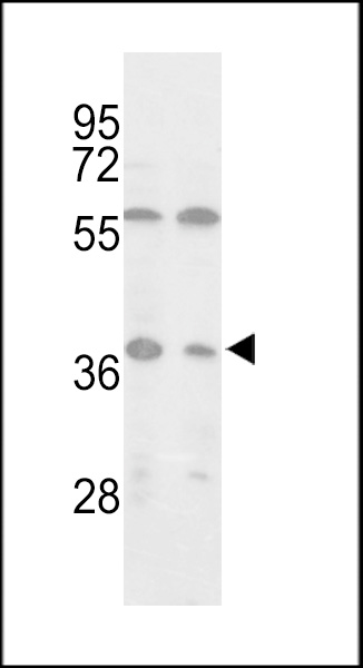
Isocitrate dehydrogenase (IDH3) Antibody (C-term)
Purified Rabbit Polyclonal Antibody (Pab)
- SPECIFICATION
- CITATIONS: 1
- PROTOCOLS
- BACKGROUND

Application
| WB, E |
|---|---|
| Primary Accession | P50213 |
| Other Accession | Q99NA5, Q9D6R2, Q28480, P41563, P56471 |
| Reactivity | Human |
| Predicted | Bovine, Monkey, Mouse, Pig, Rat |
| Host | Rabbit |
| Clonality | Polyclonal |
| Isotype | Rabbit IgG |
| Calculated MW | 39592 Da |
| Antigen Region | 317-346 aa |
| Gene ID | 3419 |
|---|---|
| Other Names | Isocitrate dehydrogenase [NAD] subunit alpha, mitochondrial, Isocitric dehydrogenase subunit alpha, NAD(+)-specific ICDH subunit alpha, IDH3A |
| Target/Specificity | This Isocitrate dehydrogenase (IDH3) antibody is generated from rabbits immunized with a KLH conjugated synthetic peptide between 317-346 amino acids from the C-terminal region of human Isocitrate dehydrogenase (IDH3). |
| Dilution | WB~~1:1000 E~~Use at an assay dependent concentration. |
| Format | Purified polyclonal antibody supplied in PBS with 0.09% (W/V) sodium azide. This antibody is prepared by Saturated Ammonium Sulfate (SAS) precipitation followed by dialysis against PBS. |
| Storage | Maintain refrigerated at 2-8°C for up to 2 weeks. For long term storage store at -20°C in small aliquots to prevent freeze-thaw cycles. |
| Precautions | Isocitrate dehydrogenase (IDH3) Antibody (C-term) is for research use only and not for use in diagnostic or therapeutic procedures. |
| Name | IDH3A (HGNC:5384) |
|---|---|
| Function | Catalytic subunit of the enzyme which catalyzes the decarboxylation of isocitrate (ICT) into alpha-ketoglutarate. The heterodimer composed of the alpha (IDH3A) and beta (IDH3B) subunits and the heterodimer composed of the alpha (IDH3A) and gamma (IDH3G) subunits, have considerable basal activity but the full activity of the heterotetramer (containing two subunits of IDH3A, one of IDH3B and one of IDH3G) requires the assembly and cooperative function of both heterodimers. |
| Cellular Location | Mitochondrion. |

Provided below are standard protocols that you may find useful for product applications.
Background
Isocitrate dehydrogenases catalyze the oxidative decarboxylation of isocitrate to 2-oxoglutarate. These enzymes belong to two distinct subclasses, one of which utilizes NAD(+) as the electron acceptor and the other NADP(+). Five isocitrate dehydrogenases have been reported: three NAD(+)-dependent isocitrate dehydrogenases, which localize to the mitochondrial matrix, and two NADP(+)-dependent isocitrate dehydrogenases, one of which is mitochondrial and the other predominantly cytosolic. NAD(+)-dependent isocitrate dehydrogenases catalyze the allosterically regulated rate-limiting step of the tricarboxylic acid cycle. Each isozyme is a heterotetramer that is composed of two alpha subunits, one beta subunit, and one gamma subunit. The protein described here is the alpha subunit of one isozyme of NAD(+)-dependent isocitrate dehydrogenase.
References
Soundar, S., et al., J. Biol. Chem. 278(52):52146-52153 (2003).
Weiss, C., et al., Biochemistry 39(7):1807-1816 (2000).
Kim, Y.O., et al., J. Biol. Chem. 274(52):36866-36875 (1999).
Huh, T.L., et al., Genomics 32(2):295-296 (1996).
Kim, Y.O., et al., Biochem. J. 308 (PT 1), 63-68 (1995) (): ().
If you have used an Abcepta product and would like to share how it has performed, please click on the "Submit Review" button and provide the requested information. Our staff will examine and post your review and contact you if needed.
If you have any additional inquiries please email technical services at tech@abcepta.com.
Ordering Information
Other Products
Shipping Information
