


 Foundational characteristics of cancer include proliferation, angiogenesis, migration, evasion of apoptosis, and cellular immortality. Find key markers for these cellular processes and antibodies to detect them.
 The SUMOplot™ Analysis Program predicts and scores sumoylation sites in your protein. SUMOylation is a post-translational modification involved in various cellular processes, such as nuclear-cytosolic transport, transcriptional regulation, apoptosis, protein stability, response to stress, and progression through the cell cycle.
 The Autophagy Receptor Motif Plotter predicts and scores autophagy receptor binding sites in your protein. Identifying proteins connected to this pathway is critical to understanding the role of autophagy in physiological as well as pathological processes such as development, differentiation, neurodegenerative diseases, stress, infection, and cancer.
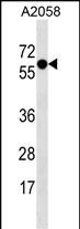
SYT4 Antibody (N-term)
Affinity Purified Rabbit Polyclonal Antibody (Pab)
- SPECIFICATION
- CITATIONS
- PROTOCOLS
- BACKGROUND

Application
| WB, E |
|---|---|
| Primary Accession | Q9H2B2 |
| Other Accession | NP_065834.1 |
| Reactivity | Human |
| Host | Rabbit |
| Clonality | Polyclonal |
| Isotype | Rabbit IgG |
| Calculated MW | 47958 Da |
| Antigen Region | 57-85 aa |
| Gene ID | 6860 |
|---|---|
| Other Names | Synaptotagmin-4, Synaptotagmin IV, SytIV, SYT4, KIAA1342 |
| Target/Specificity | This SYT4 antibody is generated from rabbits immunized with a KLH conjugated synthetic peptide between 57-85 amino acids from the N-terminal region of human SYT4. |
| Dilution | WB~~1:1000 E~~Use at an assay dependent concentration. |
| Format | Purified polyclonal antibody supplied in PBS with 0.09% (W/V) sodium azide. This antibody is purified through a protein A column, followed by peptide affinity purification. |
| Storage | Maintain refrigerated at 2-8°C for up to 2 weeks. For long term storage store at -20°C in small aliquots to prevent freeze-thaw cycles. |
| Precautions | SYT4 Antibody (N-term) is for research use only and not for use in diagnostic or therapeutic procedures. |
| Name | SYT4 |
|---|---|
| Synonyms | KIAA1342 |
| Function | Synaptotagmin family member which does not bind Ca(2+) (By similarity) (PubMed:23999003). Involved in neuronal dense core vesicles (DCVs) mobility through its interaction with KIF1A. Upon increased neuronal activity, phosphorylation by MAPK8/JNK1 destabilizes the interaction with KIF1A and captures DCVs to synapses (By similarity). Plays a role in dendrite formation by melanocytes (PubMed:23999003). |
| Cellular Location | Cytoplasmic vesicle, secretory vesicle, neuronal dense core vesicle membrane {ECO:0000250|UniProtKB:P50232}; Single-pass membrane protein {ECO:0000250|UniProtKB:P50232} |
| Tissue Location | Expressed in melanocytes (PubMed:23999003). Expressed in brain. Within brain, expression is highest in hippocampus, with substantial levels also detected in amygdala and thalamus (PubMed:23999003). |

Thousands of laboratories across the world have published research that depended on the performance of antibodies from Abcepta to advance their research. Check out links to articles that cite our products in major peer-reviewed journals, organized by research category.
info@abcepta.com, and receive a free "I Love Antibodies" mug.

Provided below are standard protocols that you may find useful for product applications.
Background
SYT4 may be involved in Ca(2+)-dependent exocytosis of secretory vesicles through Ca(2+) and phospholipid binding to the C2 domain or may serve as Ca(2+) sensors in the process of vesicular trafficking and exocytosis.
References
Need, A.C., et al. Eur. J. Hum. Genet. 17(7):946-957(2009)
Sonuga-Barke, E.J., et al. Am. J. Med. Genet. B Neuropsychiatr. Genet. 147B (8), 1359-1368 (2008) :
Ferguson, G.D., et al. J. Biol. Chem. 275(47):36920-36926(2000)
Mizutani, A., et al. J. Biol. Chem. 275(13):9823-9831(2000)
Thomas, D.M., et al. Mol. Biol. Cell 10(7):2285-2295(1999)
If you have used an Abcepta product and would like to share how it has performed, please click on the "Submit Review" button and provide the requested information. Our staff will examine and post your review and contact you if needed.
If you have any additional inquiries please email technical services at tech@abcepta.com.
Ordering Information
Shipping Information
