


 Foundational characteristics of cancer include proliferation, angiogenesis, migration, evasion of apoptosis, and cellular immortality. Find key markers for these cellular processes and antibodies to detect them.
 The SUMOplot™ Analysis Program predicts and scores sumoylation sites in your protein. SUMOylation is a post-translational modification involved in various cellular processes, such as nuclear-cytosolic transport, transcriptional regulation, apoptosis, protein stability, response to stress, and progression through the cell cycle.
 The Autophagy Receptor Motif Plotter predicts and scores autophagy receptor binding sites in your protein. Identifying proteins connected to this pathway is critical to understanding the role of autophagy in physiological as well as pathological processes such as development, differentiation, neurodegenerative diseases, stress, infection, and cancer.
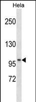
SSH1 Antibody(N-term)
Affinity Purified Rabbit Polyclonal Antibody (Pab)
- SPECIFICATION
- CITATIONS
- PROTOCOLS
- BACKGROUND

Application
| WB, E |
|---|---|
| Primary Accession | Q8WYL5 |
| Other Accession | Q76I79, NP_061857.3 |
| Reactivity | Human |
| Predicted | Mouse |
| Host | Rabbit |
| Clonality | Polyclonal |
| Isotype | Rabbit IgG |
| Calculated MW | 115511 Da |
| Antigen Region | 227-254 aa |
| Gene ID | 54434 |
|---|---|
| Other Names | Protein phosphatase Slingshot homolog 1, SSH-like protein 1, SSH-1L, hSSH-1L, SSH1, KIAA1298, SSH1L |
| Target/Specificity | This SSH1 antibody is generated from rabbits immunized with a KLH conjugated synthetic peptide between 227-254 amino acids from the N-terminal region of human SSH1. |
| Dilution | WB~~1:1000 E~~Use at an assay dependent concentration. |
| Format | Purified polyclonal antibody supplied in PBS with 0.09% (W/V) sodium azide. This antibody is purified through a protein A column, followed by peptide affinity purification. |
| Storage | Maintain refrigerated at 2-8°C for up to 2 weeks. For long term storage store at -20°C in small aliquots to prevent freeze-thaw cycles. |
| Precautions | SSH1 Antibody(N-term) is for research use only and not for use in diagnostic or therapeutic procedures. |
| Name | SSH1 (HGNC:30579) |
|---|---|
| Synonyms | KIAA1298, SSH1L |
| Function | Protein phosphatase which regulates actin filament dynamics. Dephosphorylates and activates the actin binding/depolymerizing factor cofilin, which subsequently binds to actin filaments and stimulates their disassembly. Inhibitory phosphorylation of cofilin is mediated by LIMK1, which may also be dephosphorylated and inactivated by this protein. |
| Cellular Location | Cytoplasm, cytoskeleton. Cell projection, lamellipodium. Cleavage furrow. Midbody. Note=Also recruited to actin rich membrane protrusions such as lamellipodia, which may allow local control of actin dynamics at sites of cell locomotion. Also localized to the cleavage furrow and the midbody during cytokinesis |

Thousands of laboratories across the world have published research that depended on the performance of antibodies from Abcepta to advance their research. Check out links to articles that cite our products in major peer-reviewed journals, organized by research category.
info@abcepta.com, and receive a free "I Love Antibodies" mug.

Provided below are standard protocols that you may find useful for product applications.
Background
The ADF (actin-depolymerizing factor)/cofilin family (see MIM 601442) is composed of stimulus-responsive mediators of actin dynamics. ADF/cofilin proteins are inactivated by kinases such as LIM domain kinase-1 (LIMK1; MIM 601329). The SSH family appears to play a role in actin dynamics by reactivating ADF/cofilin proteins in vivo (Niwa et al., 2002 [PubMed 11832213]).
References
Song, S.Y., et al. APMIS 118(5):389-393(2010)
Peterburs, P., et al. Cancer Res. 69(14):5634-5638(2009)
Kligys, K., et al. Biochem. Biophys. Res. Commun. 383(4):450-454(2009)
Kim, J.S., et al. Mol. Biol. Cell 20(11):2650-2660(2009)
Huang, T.Y., et al. Dev. Cell 15(5):691-703(2008)
If you have used an Abcepta product and would like to share how it has performed, please click on the "Submit Review" button and provide the requested information. Our staff will examine and post your review and contact you if needed.
If you have any additional inquiries please email technical services at tech@abcepta.com.
Ordering Information
Other Products
Shipping Information
