


 Foundational characteristics of cancer include proliferation, angiogenesis, migration, evasion of apoptosis, and cellular immortality. Find key markers for these cellular processes and antibodies to detect them.
 The SUMOplot™ Analysis Program predicts and scores sumoylation sites in your protein. SUMOylation is a post-translational modification involved in various cellular processes, such as nuclear-cytosolic transport, transcriptional regulation, apoptosis, protein stability, response to stress, and progression through the cell cycle.
 The Autophagy Receptor Motif Plotter predicts and scores autophagy receptor binding sites in your protein. Identifying proteins connected to this pathway is critical to understanding the role of autophagy in physiological as well as pathological processes such as development, differentiation, neurodegenerative diseases, stress, infection, and cancer.
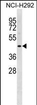
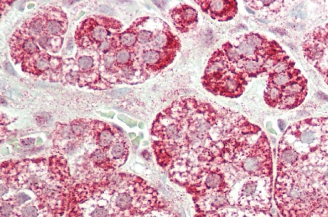
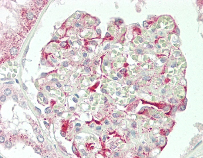
STAC2 Antibody(N-term)
Affinity Purified Rabbit Polyclonal Antibody (Pab)
- SPECIFICATION
- CITATIONS
- PROTOCOLS
- BACKGROUND

Application
| WB, IHC-P, E |
|---|---|
| Primary Accession | Q6ZMT1 |
| Other Accession | Q8R1B0, NP_945344.1 |
| Reactivity | Human |
| Predicted | Mouse |
| Host | Rabbit |
| Clonality | Polyclonal |
| Isotype | Rabbit IgG |
| Calculated MW | 45009 Da |
| Antigen Region | 1-30 aa |
| Gene ID | 342667 |
|---|---|
| Other Names | SH3 and cysteine-rich domain-containing protein 2, 24b2/STAC2, Src homology 3 and cysteine-rich domain-containing protein 2, STAC2 |
| Target/Specificity | This STAC2 antibody is generated from rabbits immunized with a KLH conjugated synthetic peptide between 1-30 amino acids from the N-terminal region of human STAC2. |
| Dilution | WB~~1:1000 IHC-P~~1:100 E~~Use at an assay dependent concentration. |
| Format | Purified polyclonal antibody supplied in PBS with 0.09% (W/V) sodium azide. This antibody is purified through a protein A column, followed by peptide affinity purification. |
| Storage | Maintain refrigerated at 2-8°C for up to 2 weeks. For long term storage store at -20°C in small aliquots to prevent freeze-thaw cycles. |
| Precautions | STAC2 Antibody(N-term) is for research use only and not for use in diagnostic or therapeutic procedures. |
| Name | STAC2 |
|---|---|
| Function | Plays a redundant role in promoting the expression of calcium channel CACNA1S at the cell membrane, and thereby contributes to increased channel activity. Slows down the inactivation rate of the calcium channel CACNA1C. |
| Cellular Location | Cytoplasm, cytosol {ECO:0000250|UniProtKB:Q8R1B0}. Cell membrane {ECO:0000250|UniProtKB:Q8R1B0}; Peripheral membrane protein {ECO:0000250|UniProtKB:Q8R1B0}; Cytoplasmic side {ECO:0000250|UniProtKB:Q8R1B0}. Cell membrane, sarcolemma {ECO:0000250|UniProtKB:Q8R1B0}; Peripheral membrane protein {ECO:0000250|UniProtKB:Q8R1B0}; Cytoplasmic side {ECO:0000250|UniProtKB:Q8R1B0}. Note=Colocalizes with CACNA1C at the plasma membrane of transfected cells. {ECO:0000250|UniProtKB:Q8R1B0} |

Thousands of laboratories across the world have published research that depended on the performance of antibodies from Abcepta to advance their research. Check out links to articles that cite our products in major peer-reviewed journals, organized by research category.
info@abcepta.com, and receive a free "I Love Antibodies" mug.

Provided below are standard protocols that you may find useful for product applications.
Background
The function of this protein is unknown.
References
Lim, J., et al. Cell 125(4):801-814(2006)
If you have used an Abcepta product and would like to share how it has performed, please click on the "Submit Review" button and provide the requested information. Our staff will examine and post your review and contact you if needed.
If you have any additional inquiries please email technical services at tech@abcepta.com.
Ordering Information
Other Products
Shipping Information
