


 Foundational characteristics of cancer include proliferation, angiogenesis, migration, evasion of apoptosis, and cellular immortality. Find key markers for these cellular processes and antibodies to detect them.
 The SUMOplot™ Analysis Program predicts and scores sumoylation sites in your protein. SUMOylation is a post-translational modification involved in various cellular processes, such as nuclear-cytosolic transport, transcriptional regulation, apoptosis, protein stability, response to stress, and progression through the cell cycle.
 The Autophagy Receptor Motif Plotter predicts and scores autophagy receptor binding sites in your protein. Identifying proteins connected to this pathway is critical to understanding the role of autophagy in physiological as well as pathological processes such as development, differentiation, neurodegenerative diseases, stress, infection, and cancer.
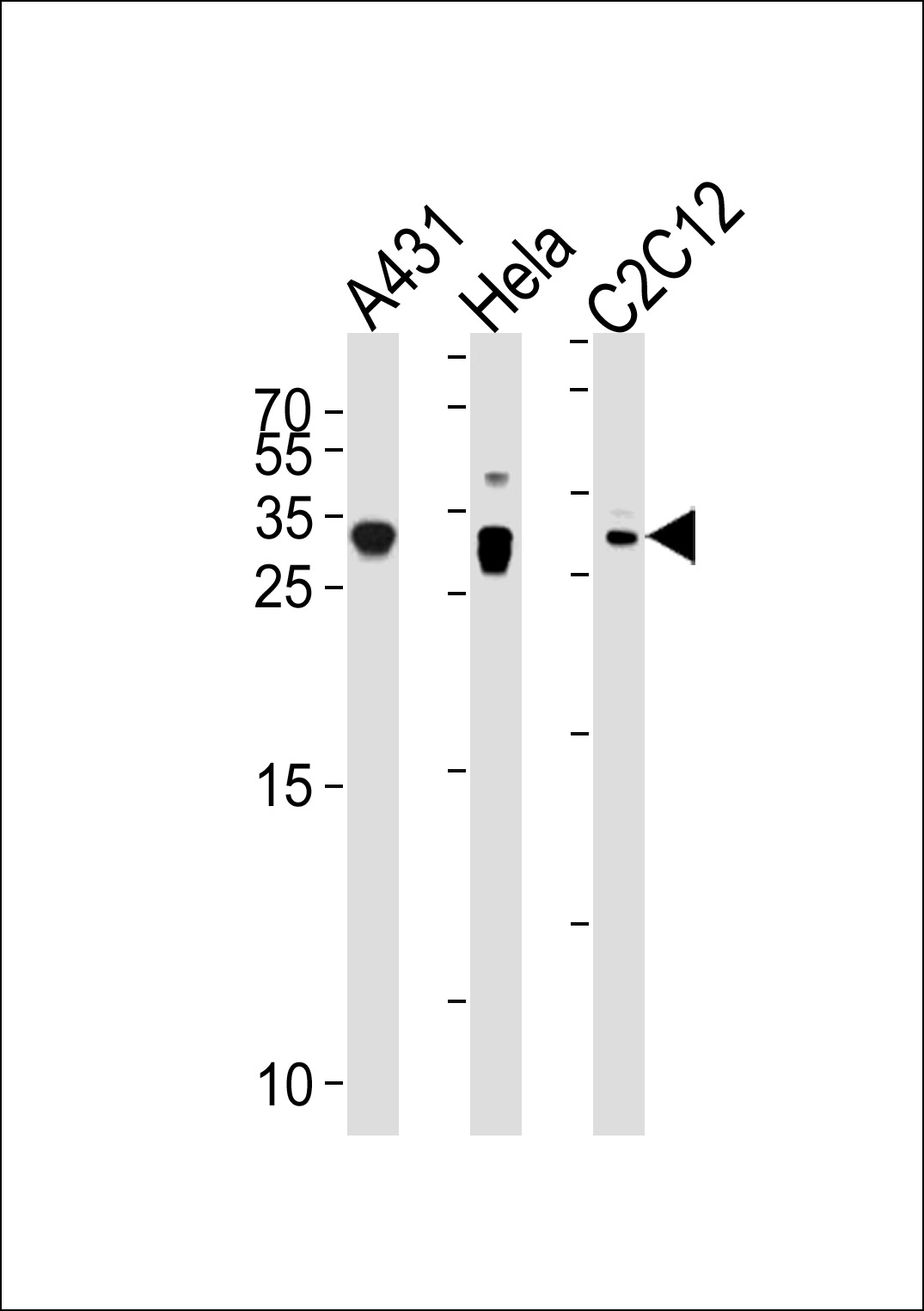
p27Kip1 Antibody (N-term S12)
Affinity Purified Rabbit Polyclonal Antibody (Pab)
- SPECIFICATION
- CITATIONS: 1
- PROTOCOLS
- BACKGROUND

Application
| WB, E |
|---|---|
| Primary Accession | P46527 |
| Other Accession | P46414, Q60439, NP_004055.1 |
| Reactivity | Human, Mouse |
| Predicted | Hamster |
| Host | Rabbit |
| Clonality | Polyclonal |
| Isotype | Rabbit IgG |
| Calculated MW | 22073 Da |
| Antigen Region | 1-30 aa |
| Gene ID | 1027 |
|---|---|
| Other Names | Cyclin-dependent kinase inhibitor 1B, Cyclin-dependent kinase inhibitor p27, p27Kip1, CDKN1B, KIP1 |
| Target/Specificity | This p27Kip1 antibody is generated from rabbits immunized with a KLH conjugated synthetic peptide between 1-30 amino acids from the N-terminal region of human p27Kip1. |
| Dilution | WB~~1:1000 E~~Use at an assay dependent concentration. |
| Format | Purified polyclonal antibody supplied in PBS with 0.09% (W/V) sodium azide. This antibody is purified through a protein A column, followed by peptide affinity purification. |
| Storage | Maintain refrigerated at 2-8°C for up to 2 weeks. For long term storage store at -20°C in small aliquots to prevent freeze-thaw cycles. |
| Precautions | p27Kip1 Antibody (N-term S12) is for research use only and not for use in diagnostic or therapeutic procedures. |
| Name | CDKN1B {ECO:0000303|PubMed:20824794} |
|---|---|
| Function | Important regulator of cell cycle progression. Inhibits the kinase activity of CDK2 bound to cyclin A, but has little inhibitory activity on CDK2 bound to SPDYA (PubMed:28666995). Involved in G1 arrest. Potent inhibitor of cyclin E- and cyclin A-CDK2 complexes. Forms a complex with cyclin type D-CDK4 complexes and is involved in the assembly, stability, and modulation of CCND1-CDK4 complex activation. Acts either as an inhibitor or an activator of cyclin type D-CDK4 complexes depending on its phosphorylation state and/or stoichometry. |
| Cellular Location | Nucleus. Cytoplasm. Endosome. Note=Nuclear and cytoplasmic in quiescent cells. AKT- or RSK-mediated phosphorylation on Thr-198, binds 14-3-3, translocates to the cytoplasm and promotes cell cycle progression. Mitogen-activated UHMK1 phosphorylation on Ser-10 also results in translocation to the cytoplasm and cell cycle progression. Phosphorylation on Ser-10 facilitates nuclear export. Translocates to the nucleus on phosphorylation of Tyr-88 and Tyr-89. Colocalizes at the endosome with SNX6; this leads to lysosomal degradation (By similarity) |
| Tissue Location | Expressed in kidney (at protein level) (PubMed:15509543). Expressed in all tissues tested (PubMed:8033212) Highest levels in skeletal muscle, lowest in liver and kidney (PubMed:8033212). |

Provided below are standard protocols that you may find useful for product applications.
Background
This gene encodes a cyclin-dependent kinase inhibitor, which shares a limited similarity with CDK inhibitor CDKN1A/p21. The encoded protein binds to and prevents the activation of cyclin E-CDK2 or cyclin D-CDK4 complexes, and thus controls the cell cycle progression at G1. The degradation of this protein, which is triggered by its CDK dependent phosphorylation and subsequent ubiquitination by SCF complexes, is required for the cellular transition from quiescence to the proliferative state. [provided by RefSeq].
References
Kajihara, R., et al. Biochem. Biophys. Res. Commun. 401(3):350-355(2010)
Kedde, M., et al. Nat. Cell Biol. 12(10):1014-1020(2010)
Canbay, E., et al. Anticancer Res. 30(7):3093-3098(2010)
Do Nascimento Borges, B., et al. In Vivo 24(4):579-582(2010)
Qin, J., et al. Hepatogastroenterology 57 (99-100), 547-553 (2010) :
If you have used an Abcepta product and would like to share how it has performed, please click on the "Submit Review" button and provide the requested information. Our staff will examine and post your review and contact you if needed.
If you have any additional inquiries please email technical services at tech@abcepta.com.
Ordering Information
Other Products
Shipping Information
