


 Foundational characteristics of cancer include proliferation, angiogenesis, migration, evasion of apoptosis, and cellular immortality. Find key markers for these cellular processes and antibodies to detect them.
 The SUMOplot™ Analysis Program predicts and scores sumoylation sites in your protein. SUMOylation is a post-translational modification involved in various cellular processes, such as nuclear-cytosolic transport, transcriptional regulation, apoptosis, protein stability, response to stress, and progression through the cell cycle.
 The Autophagy Receptor Motif Plotter predicts and scores autophagy receptor binding sites in your protein. Identifying proteins connected to this pathway is critical to understanding the role of autophagy in physiological as well as pathological processes such as development, differentiation, neurodegenerative diseases, stress, infection, and cancer.
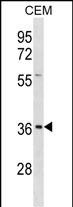
EFNB1 Antibody (Center)
Affinity Purified Rabbit Polyclonal Antibody (Pab)
- SPECIFICATION
- CITATIONS
- PROTOCOLS
- BACKGROUND

Application
| WB, E |
|---|---|
| Primary Accession | P98172 |
| Other Accession | O73612, NP_004420.1 |
| Reactivity | Human |
| Predicted | Chicken |
| Host | Rabbit |
| Clonality | Polyclonal |
| Isotype | Rabbit IgG |
| Calculated MW | 38007 Da |
| Antigen Region | 88-116 aa |
| Gene ID | 1947 |
|---|---|
| Other Names | Ephrin-B1, EFL-3, ELK ligand, ELK-L, EPH-related receptor tyrosine kinase ligand 2, LERK-2, EFNB1, EFL3, EPLG2, LERK2 |
| Target/Specificity | This EFNB1 antibody is generated from rabbits immunized with a KLH conjugated synthetic peptide between 88-116 amino acids from the Central region of human EFNB1. |
| Dilution | WB~~1:1000 E~~Use at an assay dependent concentration. |
| Format | Purified polyclonal antibody supplied in PBS with 0.09% (W/V) sodium azide. This antibody is purified through a protein A column, followed by peptide affinity purification. |
| Storage | Maintain refrigerated at 2-8°C for up to 2 weeks. For long term storage store at -20°C in small aliquots to prevent freeze-thaw cycles. |
| Precautions | EFNB1 Antibody (Center) is for research use only and not for use in diagnostic or therapeutic procedures. |
| Name | EFNB1 |
|---|---|
| Synonyms | EFL3, EPLG2, LERK2 |
| Function | Cell surface transmembrane ligand for Eph receptors, a family of receptor tyrosine kinases which are crucial for migration, repulsion and adhesion during neuronal, vascular and epithelial development (PubMed:7973638, PubMed:8070404). Binding to Eph receptors residing on adjacent cells leads to contact-dependent bidirectional signaling into neighboring cells (PubMed:7973638, PubMed:8070404). Shows high affinity for the receptor tyrosine kinase EPHB1/ELK (PubMed:7973638, PubMed:8070404). Can also bind EPHB2 and EPHB3 (PubMed:8070404). Binds to, and induces collapse of, commissural axons/growth cones in vitro (By similarity). May play a role in constraining the orientation of longitudinally projecting axons (By similarity). |
| Cellular Location | Cell membrane; Single-pass type I membrane protein. Membrane raft. Note=May recruit GRIP1 and GRIP2 to membrane raft domains [Ephrin-B1 intracellular domain]: Nucleus. Note=Colocalizes with ZHX2 in the nucleus. {ECO:0000250|UniProtKB:P52795} |
| Tissue Location | Widely expressed (PubMed:7973638, PubMed:8070404). Detected in both neuronal and non-neuronal tissues (PubMed:7973638, PubMed:8070404). Seems to have particularly strong expression in retina, sciatic nerve, heart and spinal cord (PubMed:7973638) |

Thousands of laboratories across the world have published research that depended on the performance of antibodies from Abcepta to advance their research. Check out links to articles that cite our products in major peer-reviewed journals, organized by research category.
info@abcepta.com, and receive a free "I Love Antibodies" mug.

Provided below are standard protocols that you may find useful for product applications.
Background
The protein encoded by this gene is a type I membrane protein and a ligand of Eph-related receptor tyrosine kinases. It may play a role in cell adhesion and function in the development or maintenance of the nervous system.
References
Hogue, J., et al. Am. J. Med. Genet. A 152A (10), 2574-2577 (2010) :
Arvanitis, D.N., et al. Mol. Cell. Biol. 30(10):2508-2517(2010)
Makarov, R., et al. BMC Med. Genet. 11, 98 (2010) :
Vazin, T., et al. PLoS ONE 4 (8), E6606 (2009) :
Wallis, D., et al. Am. J. Med. Genet. A 146A (15), 2008-2012 (2008) :
If you have used an Abcepta product and would like to share how it has performed, please click on the "Submit Review" button and provide the requested information. Our staff will examine and post your review and contact you if needed.
If you have any additional inquiries please email technical services at tech@abcepta.com.
Ordering Information
Other Products
Shipping Information
