


 Foundational characteristics of cancer include proliferation, angiogenesis, migration, evasion of apoptosis, and cellular immortality. Find key markers for these cellular processes and antibodies to detect them.
 The SUMOplot™ Analysis Program predicts and scores sumoylation sites in your protein. SUMOylation is a post-translational modification involved in various cellular processes, such as nuclear-cytosolic transport, transcriptional regulation, apoptosis, protein stability, response to stress, and progression through the cell cycle.
 The Autophagy Receptor Motif Plotter predicts and scores autophagy receptor binding sites in your protein. Identifying proteins connected to this pathway is critical to understanding the role of autophagy in physiological as well as pathological processes such as development, differentiation, neurodegenerative diseases, stress, infection, and cancer.
POLR2G Antibody (C-term)
Affinity Purified Rabbit Polyclonal Antibody (Pab)
- SPECIFICATION
- CITATIONS
- PROTOCOLS
- BACKGROUND

Application
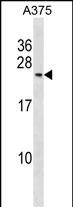
| WB, E |
|---|---|
| Primary Accession | P62487 |
| Other Accession | P62489, P62488, Q7ZW41, Q5E9B8, NP_002687.1 |
| Reactivity | Human |
| Predicted | Bovine, Zebrafish, Mouse, Rat |
| Host | Rabbit |
| Clonality | Polyclonal |
| Isotype | Rabbit IgG |
| Calculated MW | 19294 Da |
| Antigen Region | 125-154 aa |
| Gene ID | 5436 |
|---|---|
| Other Names | DNA-directed RNA polymerase II subunit RPB7, RNA polymerase II subunit B7, DNA-directed RNA polymerase II subunit G, RNA polymerase II 19 kDa subunit, RPB19, POLR2G, RPB7 |
| Target/Specificity | This POLR2G antibody is generated from rabbits immunized with a KLH conjugated synthetic peptide between 125-154 amino acids from the C-terminal region of human POLR2G. |
| Dilution | WB~~1:1000 E~~Use at an assay dependent concentration. |
| Format | Purified polyclonal antibody supplied in PBS with 0.09% (W/V) sodium azide. This antibody is purified through a protein A column, followed by peptide affinity purification. |
| Storage | Maintain refrigerated at 2-8°C for up to 2 weeks. For long term storage store at -20°C in small aliquots to prevent freeze-thaw cycles. |
| Precautions | POLR2G Antibody (C-term) is for research use only and not for use in diagnostic or therapeutic procedures. |
| Name | POLR2G |
|---|---|
| Synonyms | RPB7 |
| Function | Core component of RNA polymerase II (Pol II), a DNA-dependent RNA polymerase which synthesizes mRNA precursors and many functional non-coding RNAs using the four ribonucleoside triphosphates as substrates. Pol II is the central component of the basal RNA polymerase II transcription machinery. It is composed of mobile elements that move relative to each other. POLR2G/RPB7 is part of a subcomplex with POLR2D/RPB4 that binds to a pocket formed by POLR2A/RPB1, POLR2B/RPB2 and POLR2F/RPABC2 at the base of the clamp element. The POLR2D/RPB4- POLR2G/RPB7 subcomplex seems to lock the clamp via POLR2G/RPB7 in the closed conformation thus preventing double-stranded DNA to enter the active site cleft. The POLR2D/RPB4-POLR2G/RPB7 subcomplex binds single- stranded DNA and RNA. |
| Cellular Location | Nucleus. |

Thousands of laboratories across the world have published research that depended on the performance of antibodies from Abcepta to advance their research. Check out links to articles that cite our products in major peer-reviewed journals, organized by research category.
info@abcepta.com, and receive a free "I Love Antibodies" mug.

Provided below are standard protocols that you may find useful for product applications.
Background
This gene encodes the seventh largest subunit of RNA polymerase II, the polymerase responsible for synthesizing messenger RNA in eukaryotes. The protein functions in transcription initiation, and is also thought to help stabilize transcribing polyermase molecules during elongation.
References
Cojocaru, M., et al. Biochem. J. 409(1):139-147(2008)
Ujvari, A., et al. Nat. Struct. Mol. Biol. 13(1):49-54(2006)
Meka, H., et al. Nucleic Acids Res. 33(19):6435-6444(2005)
Lehner, B., et al. Genomics 83(1):153-167(2004)
Zhou, M., et al. Proc. Natl. Acad. Sci. U.S.A. 100(22):12666-12671(2003)
If you have used an Abcepta product and would like to share how it has performed, please click on the "Submit Review" button and provide the requested information. Our staff will examine and post your review and contact you if needed.
If you have any additional inquiries please email technical services at tech@abcepta.com.
Ordering Information
Other Products
Shipping Information
