


 Foundational characteristics of cancer include proliferation, angiogenesis, migration, evasion of apoptosis, and cellular immortality. Find key markers for these cellular processes and antibodies to detect them.
 The SUMOplot™ Analysis Program predicts and scores sumoylation sites in your protein. SUMOylation is a post-translational modification involved in various cellular processes, such as nuclear-cytosolic transport, transcriptional regulation, apoptosis, protein stability, response to stress, and progression through the cell cycle.
 The Autophagy Receptor Motif Plotter predicts and scores autophagy receptor binding sites in your protein. Identifying proteins connected to this pathway is critical to understanding the role of autophagy in physiological as well as pathological processes such as development, differentiation, neurodegenerative diseases, stress, infection, and cancer.
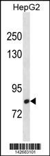
CMIP Antibody (C-term)
Affinity Purified Rabbit Polyclonal Antibody (Pab)
- SPECIFICATION
- CITATIONS
- PROTOCOLS
- BACKGROUND

Application
| WB, E |
|---|---|
| Primary Accession | Q8IY22 |
| Other Accession | A1L3F5, Q9D486, NP_085132.1 |
| Reactivity | Human |
| Predicted | Mouse, Xenopus |
| Host | Rabbit |
| Clonality | Polyclonal |
| Isotype | Rabbit IgG |
| Calculated MW | 86331 Da |
| Antigen Region | 735-764 aa |
| Gene ID | 80790 |
|---|---|
| Other Names | C-Maf-inducing protein, c-Mip, Truncated c-Maf-inducing protein, Tc-Mip, CMIP, KIAA1694, TCMIP |
| Target/Specificity | This CMIP antibody is generated from rabbits immunized with a KLH conjugated synthetic peptide between 735-764 amino acids from the C-terminal region of human CMIP. |
| Dilution | WB~~1:1000 E~~Use at an assay dependent concentration. |
| Format | Purified polyclonal antibody supplied in PBS with 0.09% (W/V) sodium azide. This antibody is purified through a protein A column, followed by peptide affinity purification. |
| Storage | Maintain refrigerated at 2-8°C for up to 2 weeks. For long term storage store at -20°C in small aliquots to prevent freeze-thaw cycles. |
| Precautions | CMIP Antibody (C-term) is for research use only and not for use in diagnostic or therapeutic procedures. |
| Name | CMIP |
|---|---|
| Synonyms | KIAA1694, TCMIP |
| Function | Plays a role in T-cell signaling pathway. Isoform 2 may play a role in T-helper 2 (Th2) signaling pathway and seems to represent the first proximal signaling protein that links T-cell receptor-mediated signal to the activation of c-Maf Th2 specific factor. |
| Cellular Location | Nucleus. Cytoplasm. Note=Isoform 2 is translocated to the nucleus and is specifically recruited during minimal change nephrotic syndrome (MCNS) (PubMed:12939343, PubMed:15616553). Detected in nuclear and cytoplasmic compartments during MCNS relapse (PubMed:12939343, PubMed:15616553). Expressed in cytoplasm only during MCNS remission and absent in normal patients (PubMed:12939343) |
| Tissue Location | Isoform 1 is expressed in peripheral blood mononuclear cells and kidney. Lower expression in brain and liver Expression is down-regulated in activated cells. Isoform 2 is expressed in lymphocyte precursors, however, expression shuts down during maturation and differentiation in thymus and fetal liver |

Thousands of laboratories across the world have published research that depended on the performance of antibodies from Abcepta to advance their research. Check out links to articles that cite our products in major peer-reviewed journals, organized by research category.
info@abcepta.com, and receive a free "I Love Antibodies" mug.

Provided below are standard protocols that you may find useful for product applications.
Background
CMIP plays a role in T-cell signaling pathway. Isoform 2 may play a role in T-helper 2 (Th2) signaling pathway and seems to represent the first proximal signaling protein that links T-cell receptor-mediated signal to the activation of c-Maf Th2 specific factor.
References
Rose, J.E., et al. Mol. Med. 16 (7-8), 247-253 (2010) :
Audard, V., et al. Blood 115(18):3756-3762(2010)
Kamal, M., et al. FEBS Lett. 584(3):500-506(2010)
Zhang, S.Y., et al. Sci Signal 3 (122), RA39 (2010) :
Newbury, D.F., et al. Am. J. Hum. Genet. 85(2):264-272(2009)
If you have used an Abcepta product and would like to share how it has performed, please click on the "Submit Review" button and provide the requested information. Our staff will examine and post your review and contact you if needed.
If you have any additional inquiries please email technical services at tech@abcepta.com.
Ordering Information
Other Products
Shipping Information
