


 Foundational characteristics of cancer include proliferation, angiogenesis, migration, evasion of apoptosis, and cellular immortality. Find key markers for these cellular processes and antibodies to detect them.
 The SUMOplot™ Analysis Program predicts and scores sumoylation sites in your protein. SUMOylation is a post-translational modification involved in various cellular processes, such as nuclear-cytosolic transport, transcriptional regulation, apoptosis, protein stability, response to stress, and progression through the cell cycle.
 The Autophagy Receptor Motif Plotter predicts and scores autophagy receptor binding sites in your protein. Identifying proteins connected to this pathway is critical to understanding the role of autophagy in physiological as well as pathological processes such as development, differentiation, neurodegenerative diseases, stress, infection, and cancer.
> home > Products > Primary Antibodies > Antibody Collections > SARS-Human interaction partners > CCDC155 Antibody (Center)
CCDC155 Antibody (Center)
Affinity Purified Rabbit Polyclonal Antibody (Pab)
- SPECIFICATION
- CITATIONS
- PROTOCOLS
- BACKGROUND

Application
| WB, E |
|---|---|
| Primary Accession | Q8N6L0 |
| Reactivity | Human |
| Host | Rabbit |
| Clonality | Polyclonal |
| Isotype | Rabbit IgG |
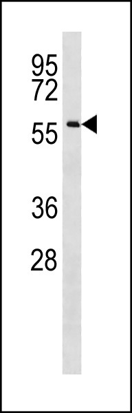
| Calculated MW | 62783 Da |
| Antigen Region | 159-185 aa |
| Gene ID | 147872 |
|---|---|
| Other Names | Protein KASH5, Coiled-coil domain-containing protein 155, KASH domain-containing protein 5, CCDC155, KASH5 |
| Target/Specificity | This CCDC155 antibody is generated from rabbits immunized with a KLH conjugated synthetic peptide between 159-185 amino acids from the Central region of human CCDC155. |
| Dilution | WB~~1:1000 E~~Use at an assay dependent concentration. |
| Format | Purified polyclonal antibody supplied in PBS with 0.09% (W/V) sodium azide. This antibody is purified through a protein A column, followed by peptide affinity purification. |
| Storage | Maintain refrigerated at 2-8°C for up to 2 weeks. For long term storage store at -20°C in small aliquots to prevent freeze-thaw cycles. |
| Precautions | CCDC155 Antibody (Center) is for research use only and not for use in diagnostic or therapeutic procedures. |
| Name | KASH5 (HGNC:26520) |
|---|---|
| Synonyms | CCDC155 |
| Function | As a component of the LINC (LInker of Nucleoskeleton and Cytoskeleton) complex, involved in the connection between the nuclear lamina and the cytoskeleton. The nucleocytoplasmic interactions established by the LINC complex play an important role in the transmission of mechanical forces across the nuclear envelope and in nuclear movement and positioning. Required for telomere attachment to nuclear envelope in the prophase of meiosis (PubMed:35587281). Required for rapid telomere prophase movements implicating a SUN1/2:KASH5 LINC complex in which SUN1 and SUN2 seem to act at least partial redundantly. Required for homolog pairing during meiotic prophase in spermatocytes and probably oocytes. Essential for male and female gametogenesis (PubMed:35587281). Recruits cytoplasmic dynein to telomere attachment sites at the nuclear envelope in spermatocytes. In oocytes is involved in meiotic resumption and spindle formation. |
| Cellular Location | Nucleus outer membrane {ECO:0000250|UniProtKB:Q80VJ8, ECO:0000305}; Single-pass type IV membrane protein; Cytoplasmic side. Nucleus {ECO:0000250|UniProtKB:Q80VJ8}. Chromosome, telomere {ECO:0000250|UniProtKB:Q80VJ8}. Nucleus envelope. Note=Localized exclusively at telomeres from the leptotene to diplotene stages. Colocalizes with SUN2 at sites of telomere attachment in meiocytes. At oocyte MI stage localized around the spindle, at MII stage localized to the spindle poles {ECO:0000250|UniProtKB:Q80VJ8} |
| Tissue Location | Expressed in testis (at protein level). |
Research Areas
Citations (0)

Thousands of laboratories across the world have published research that depended on the performance of antibodies from Abcepta to advance their research. Check out links to articles that cite our products in major peer-reviewed journals, organized by research category.
Submit your citation using an Abcepta antibody to
info@abcepta.com, and receive a free "I Love Antibodies" mug.
info@abcepta.com, and receive a free "I Love Antibodies" mug.

Application Protocols
Provided below are standard protocols that you may find useful for product applications.
Background
The function of this protein remains unknown.
Abcepta welcomes feedback from its customers.
If you have used an Abcepta product and would like to share how it has performed, please click on the "Submit Review" button and provide the requested information. Our staff will examine and post your review and contact you if needed.
If you have any additional inquiries please email technical services at tech@abcepta.com.
$ 150.00
$ 385.00
Cat# AP20386c
Ordering Information
United States
AlbaniaAustraliaAustriaBelgiumBosnia & HerzegovinaBrazilBulgariaCanadaCentral AmericaChinaCroatiaCyprusCzech RepublicDenmarkEstoniaFinlandFranceGermanyGreeceHong KongHungaryIcelandIndiaIndonesiaIrelandIsraelItalyJapanLatviaLithuaniaLuxembourgMacedoniaMalaysiaMaltaMexicoNetherlandsNew ZealandNorwayPakistanPolandPortugalRomaniaSerbiaSingaporeSlovakiaSloveniaSouth AfricaSouth KoreaSpainSwedenSwitzerlandTaiwanTurkeyUnited KingdomUnited StatesVietnamWorldwideOthers
USA Headquarters
(888) 735-7227 / (858) 622-0099 or (858) 875-1900
Other Products
Shipping Information
Domestic orders (in stock items)
Shipped out the same day. Orders placed after 1 PM (PST) will ship out the next business day.
International orders
Contact your local distributors
