


 Foundational characteristics of cancer include proliferation, angiogenesis, migration, evasion of apoptosis, and cellular immortality. Find key markers for these cellular processes and antibodies to detect them.
 The SUMOplot™ Analysis Program predicts and scores sumoylation sites in your protein. SUMOylation is a post-translational modification involved in various cellular processes, such as nuclear-cytosolic transport, transcriptional regulation, apoptosis, protein stability, response to stress, and progression through the cell cycle.
 The Autophagy Receptor Motif Plotter predicts and scores autophagy receptor binding sites in your protein. Identifying proteins connected to this pathway is critical to understanding the role of autophagy in physiological as well as pathological processes such as development, differentiation, neurodegenerative diseases, stress, infection, and cancer.
> home > Products > Primary Antibodies > Antibody Collections > Catalog Updated > MORC2 Antibody (C-term)
MORC2 Antibody (C-term)
Purified Rabbit Polyclonal Antibody (Pab)
- SPECIFICATION
- CITATIONS: 1
- PROTOCOLS
- BACKGROUND

Application
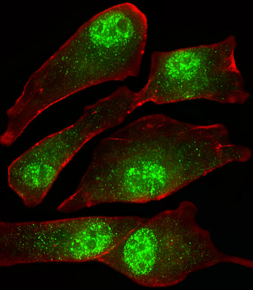
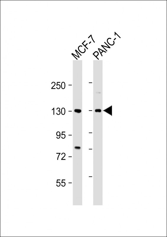
| IF, WB, E |
|---|---|
| Primary Accession | Q9Y6X9 |
| Reactivity | Human |
| Host | Rabbit |
| Clonality | Polyclonal |
| Isotype | Rabbit IgG |
| Calculated MW | 117823 Da |
| Antigen Region | 828-862 aa |
| Gene ID | 22880 |
|---|---|
| Other Names | MORC family CW-type zinc finger protein 2, Zinc finger CW-type coiled-coil domain protein 1, MORC2, KIAA0852, ZCWCC1 |
| Target/Specificity | This MORC2 antibody is generated from a rabbit immunized with a KLH conjugated synthetic peptide between 828-862 amino acids from the C-terminal region of human MORC2. |
| Dilution | IF~~1:25 WB~~1:2000 E~~Use at an assay dependent concentration. |
| Format | Purified polyclonal antibody supplied in PBS with 0.09% (W/V) sodium azide. This antibody is purified through a protein A column, followed by peptide affinity purification. |
| Storage | Maintain refrigerated at 2-8°C for up to 2 weeks. For long term storage store at -20°C in small aliquots to prevent freeze-thaw cycles. |
| Precautions | MORC2 Antibody (C-term) is for research use only and not for use in diagnostic or therapeutic procedures. |
| Name | MORC2 (HGNC:23573) |
|---|---|
| Synonyms | KIAA0852, ZCWCC1 |
| Function | Essential for epigenetic silencing by the HUSH (human silencing hub) complex. Recruited by HUSH to target site in heterochromatin, the ATPase activity and homodimerization are critical for HUSH-mediated silencing (PubMed:28581500, PubMed:29440755, PubMed:32693025). Represses germ cell-related genes and L1 retrotransposons in collaboration with SETDB1 and the HUSH complex, the silencing is dependent of repressive epigenetic modifications, such as H3K9me3 mark. Silencing events often occur within introns of transcriptionally active genes, and lead to the down-regulation of host gene expression (PubMed:29211708). During DNA damage response, regulates chromatin remodeling through ATP hydrolysis. Upon DNA damage, is phosphorylated by PAK1, both colocalize to chromatin and induce H2AX expression. ATPase activity is required and dependent of phosphorylation by PAK1 and presence of DNA (PubMed:23260667). Recruits histone deacetylases, such as HDAC4, to promoter regions, causing local histone H3 deacetylation and transcriptional repression of genes such as CA9 (PubMed:20110259, PubMed:20225202). Exhibits a cytosolic function in lipogenesis, adipogenic differentiation, and lipid homeostasis by increasing the activity of ACLY, possibly preventing its dephosphorylation (PubMed:24286864). |
| Cellular Location | Nucleus. Cytoplasm, cytosol Chromosome Nucleus matrix. Note=Mainly located in the nucleus (PubMed:20225202). Upon phosphorylation at Ser-739, recruited to damaged chromatin (PubMed:23260667) |
| Tissue Location | Highly expressed in smooth muscle, pancreas and testis |
Research Areas
Citations ( 0 )

Application Protocols
Provided below are standard protocols that you may find useful for product applications.
Background
May act as a transcriptional repressor. Down-regulates CA9 expression.
References
Nagase T.,et al.DNA Res. 5:355-364(1998).
Collins J.E.,et al.Genome Biol. 5:R84.1-R84.11(2004).
Dunham I.,et al.Nature 402:489-495(1999).
Mural R.J.,et al.Submitted (JUL-2005) to the EMBL/GenBank/DDBJ databases.
Bechtel S.,et al.BMC Genomics 8:399-399(2007).
Abcepta welcomes feedback from its customers.
If you have used an Abcepta product and would like to share how it has performed, please click on the "Submit Review" button and provide the requested information. Our staff will examine and post your review and contact you if needed.
If you have any additional inquiries please email technical services at tech@abcepta.com.
$ 150.00
$ 385.00
Cat# AP20588c
Ordering Information
United States
AlbaniaAustraliaAustriaBelgiumBosnia & HerzegovinaBrazilBulgariaCanadaCentral AmericaChinaCroatiaCyprusCzech RepublicDenmarkEstoniaFinlandFranceGermanyGreeceHong KongHungaryIcelandIndiaIndonesiaIrelandIsraelItalyJapanLatviaLithuaniaLuxembourgMacedoniaMalaysiaMaltaMexicoNetherlandsNew ZealandNorwayPakistanPolandPortugalRomaniaSerbiaSingaporeSlovakiaSloveniaSouth AfricaSouth KoreaSpainSwedenSwitzerlandTaiwanTurkeyUnited KingdomUnited StatesVietnamWorldwideOthers
USA Headquarters
(888) 735-7227 / (858) 622-0099 or (858) 875-1900
Other Products
Shipping Information
Domestic orders (in stock items)
Shipped out the same day. Orders placed after 1 PM (PST) will ship out the next business day.
International orders
Contact your local distributors
