


 Foundational characteristics of cancer include proliferation, angiogenesis, migration, evasion of apoptosis, and cellular immortality. Find key markers for these cellular processes and antibodies to detect them.
 The SUMOplot™ Analysis Program predicts and scores sumoylation sites in your protein. SUMOylation is a post-translational modification involved in various cellular processes, such as nuclear-cytosolic transport, transcriptional regulation, apoptosis, protein stability, response to stress, and progression through the cell cycle.
 The Autophagy Receptor Motif Plotter predicts and scores autophagy receptor binding sites in your protein. Identifying proteins connected to this pathway is critical to understanding the role of autophagy in physiological as well as pathological processes such as development, differentiation, neurodegenerative diseases, stress, infection, and cancer.
CPLX2 Antibody (Center)
Purified Rabbit Polyclonal Antibody (Pab)
- SPECIFICATION
- CITATIONS
- PROTOCOLS
- BACKGROUND

Application
| WB, E |
|---|---|
| Primary Accession | Q6PUV4 |
| Other Accession | P84087, P84086, P84088 |
| Reactivity | Mouse |
| Predicted | Bovine, Rat |
| Host | Rabbit |
| Clonality | Polyclonal |
| Isotype | Rabbit IgG |
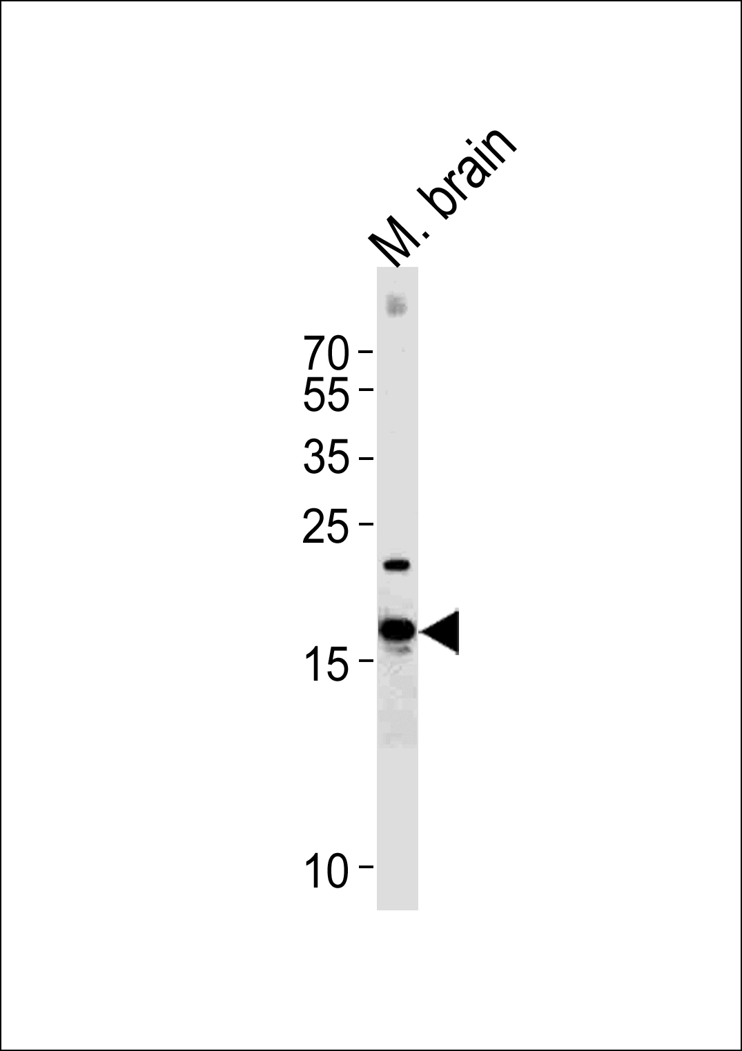
| Calculated MW | 15394 Da |
| Gene ID | 10814 |
|---|---|
| Other Names | Complexin-2, Complexin II, CPX II, Synaphin-1, CPLX2 |
| Target/Specificity | This CPLX2 antibody is generated from a rabbit immunized with a KLH conjugated synthetic peptide between 51-83 amino acids from the Central region of human CPLX2. |
| Dilution | WB~~1:1000 E~~Use at an assay dependent concentration. |
| Format | Purified polyclonal antibody supplied in PBS with 0.09% (W/V) sodium azide. This antibody is purified through a protein A column, followed by peptide affinity purification. |
| Storage | Maintain refrigerated at 2-8°C for up to 2 weeks. For long term storage store at -20°C in small aliquots to prevent freeze-thaw cycles. |
| Precautions | CPLX2 Antibody (Center) is for research use only and not for use in diagnostic or therapeutic procedures. |
| Name | CPLX2 |
|---|---|
| Function | Negatively regulates the formation of synaptic vesicle clustering at active zone to the presynaptic membrane in postmitotic neurons. Positively regulates a late step in exocytosis of various cytoplasmic vesicles, such as synaptic vesicles and other secretory vesicles. Also involved in mast cell exocytosis (By similarity). |
| Cellular Location | Cytoplasm, cytosol {ECO:0000250|UniProtKB:P84087}. Presynapse {ECO:0000250|UniProtKB:P84087}. Nucleus {ECO:0000250|UniProtKB:P84087} Perikaryon {ECO:0000250|UniProtKB:P84087}. Note=Translocated from the perikaryon to the presynaptic terminals during maturation of neuronal cells. In mast cells, cytosol and nucleus. Becomes enriched near plasma membrane following stimulation. {ECO:0000250|UniProtKB:P84087} |
| Tissue Location | Nervous system. In hippocampus and cerebellum, expressed mainly by excitatory neurons. Down-regulated in brain cortex from patients suffering from Huntington disease, bipolar disorder or major depression. Down-regulated in cerebellum from patients with schizophrenia. |

Thousands of laboratories across the world have published research that depended on the performance of antibodies from Abcepta to advance their research. Check out links to articles that cite our products in major peer-reviewed journals, organized by research category.
info@abcepta.com, and receive a free "I Love Antibodies" mug.

Provided below are standard protocols that you may find useful for product applications.
Background
Negatively regulates the formation of synaptic vesicle clustering at active zone to the presynaptic membrane in postmitotic neurons. Positively regulates a late step in synaptic vesicle exocytosis. Also involved in mast cell exocytosis (By similarity).
References
McMahon H.T.,et al.Cell 83:111-119(1995).
Ota T.,et al.Nat. Genet. 36:40-45(2004).
Raevskaya N.M.,et al.Gene 359:127-137(2005).
Harrison P.J.,et al.Lancet 352:1669-1673(1998).
Eastwood S.L.,et al.Brain Res. Bull. 55:569-578(2001).
If you have used an Abcepta product and would like to share how it has performed, please click on the "Submit Review" button and provide the requested information. Our staff will examine and post your review and contact you if needed.
If you have any additional inquiries please email technical services at tech@abcepta.com.
Ordering Information
Shipping Information
