


 Foundational characteristics of cancer include proliferation, angiogenesis, migration, evasion of apoptosis, and cellular immortality. Find key markers for these cellular processes and antibodies to detect them.
 The SUMOplot™ Analysis Program predicts and scores sumoylation sites in your protein. SUMOylation is a post-translational modification involved in various cellular processes, such as nuclear-cytosolic transport, transcriptional regulation, apoptosis, protein stability, response to stress, and progression through the cell cycle.
 The Autophagy Receptor Motif Plotter predicts and scores autophagy receptor binding sites in your protein. Identifying proteins connected to this pathway is critical to understanding the role of autophagy in physiological as well as pathological processes such as development, differentiation, neurodegenerative diseases, stress, infection, and cancer.
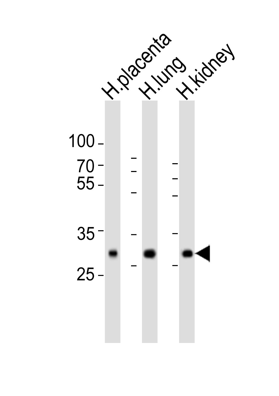
SOD3 Antibody (C-term)
Purified Rabbit Polyclonal Antibody (Pab)
- SPECIFICATION
- CITATIONS
- PROTOCOLS
- BACKGROUND

Application
| WB, E |
|---|---|
| Primary Accession | P08294 |
| Reactivity | Human |
| Host | Rabbit |
| Clonality | Polyclonal |
| Isotype | Rabbit IgG |
| Calculated MW | 25851 Da |
| Gene ID | 6649 |
|---|---|
| Other Names | Extracellular superoxide dismutase [Cu-Zn], EC-SOD, SOD3 |
| Target/Specificity | This SOD3 antibody is generated from a rabbit immunized with a KLH conjugated synthetic peptide between 186-219 amino acids from the C-terminal region of human SOD3. |
| Dilution | WB~~1:500-1:1000 E~~Use at an assay dependent concentration. |
| Format | Purified polyclonal antibody supplied in PBS with 0.09% (W/V) sodium azide. This antibody is purified through a protein A column, followed by peptide affinity purification. |
| Storage | Maintain refrigerated at 2-8°C for up to 2 weeks. For long term storage store at -20°C in small aliquots to prevent freeze-thaw cycles. |
| Precautions | SOD3 Antibody (C-term) is for research use only and not for use in diagnostic or therapeutic procedures. |
| Name | SOD3 |
|---|---|
| Function | Protect the extracellular space from toxic effect of reactive oxygen intermediates by converting superoxide radicals into hydrogen peroxide and oxygen. |
| Cellular Location | Secreted, extracellular space. Golgi apparatus, trans-Golgi network {ECO:0000250|UniProtKB:O09164}. Note=99% of EC-SOD is anchored to heparan sulfate proteoglycans in the tissue interstitium, and 1% is located in the vasculature in equilibrium between the plasma and the endothelium |
| Tissue Location | Expressed in blood vessels, heart, lung, kidney and placenta. Major SOD isoenzyme in extracellular fluids such as plasma, lymph and synovial fluid |

Thousands of laboratories across the world have published research that depended on the performance of antibodies from Abcepta to advance their research. Check out links to articles that cite our products in major peer-reviewed journals, organized by research category.
info@abcepta.com, and receive a free "I Love Antibodies" mug.

Provided below are standard protocols that you may find useful for product applications.
Background
Protect the extracellular space from toxic effect of reactive oxygen intermediates by converting superoxide radicals into hydrogen peroxide and oxygen.
References
Hjalmarsson K.,et al.Proc. Natl. Acad. Sci. U.S.A. 84:6340-6344(1987).
Folz R.J.,et al.Genomics 22:162-171(1994).
Halleck A.,et al.Submitted (JUN-2004) to the EMBL/GenBank/DDBJ databases.
Adachi T.,et al.Free Radic. Biol. Med. 13:205-210(1992).
Nozik-Grayck E.,et al.Int. J. Biochem. Cell Biol. 37:2466-2471(2005).
If you have used an Abcepta product and would like to share how it has performed, please click on the "Submit Review" button and provide the requested information. Our staff will examine and post your review and contact you if needed.
If you have any additional inquiries please email technical services at tech@abcepta.com.

Ordering Information
Other Products
Shipping Information
