


 Foundational characteristics of cancer include proliferation, angiogenesis, migration, evasion of apoptosis, and cellular immortality. Find key markers for these cellular processes and antibodies to detect them.
 The SUMOplot™ Analysis Program predicts and scores sumoylation sites in your protein. SUMOylation is a post-translational modification involved in various cellular processes, such as nuclear-cytosolic transport, transcriptional regulation, apoptosis, protein stability, response to stress, and progression through the cell cycle.
 The Autophagy Receptor Motif Plotter predicts and scores autophagy receptor binding sites in your protein. Identifying proteins connected to this pathway is critical to understanding the role of autophagy in physiological as well as pathological processes such as development, differentiation, neurodegenerative diseases, stress, infection, and cancer.
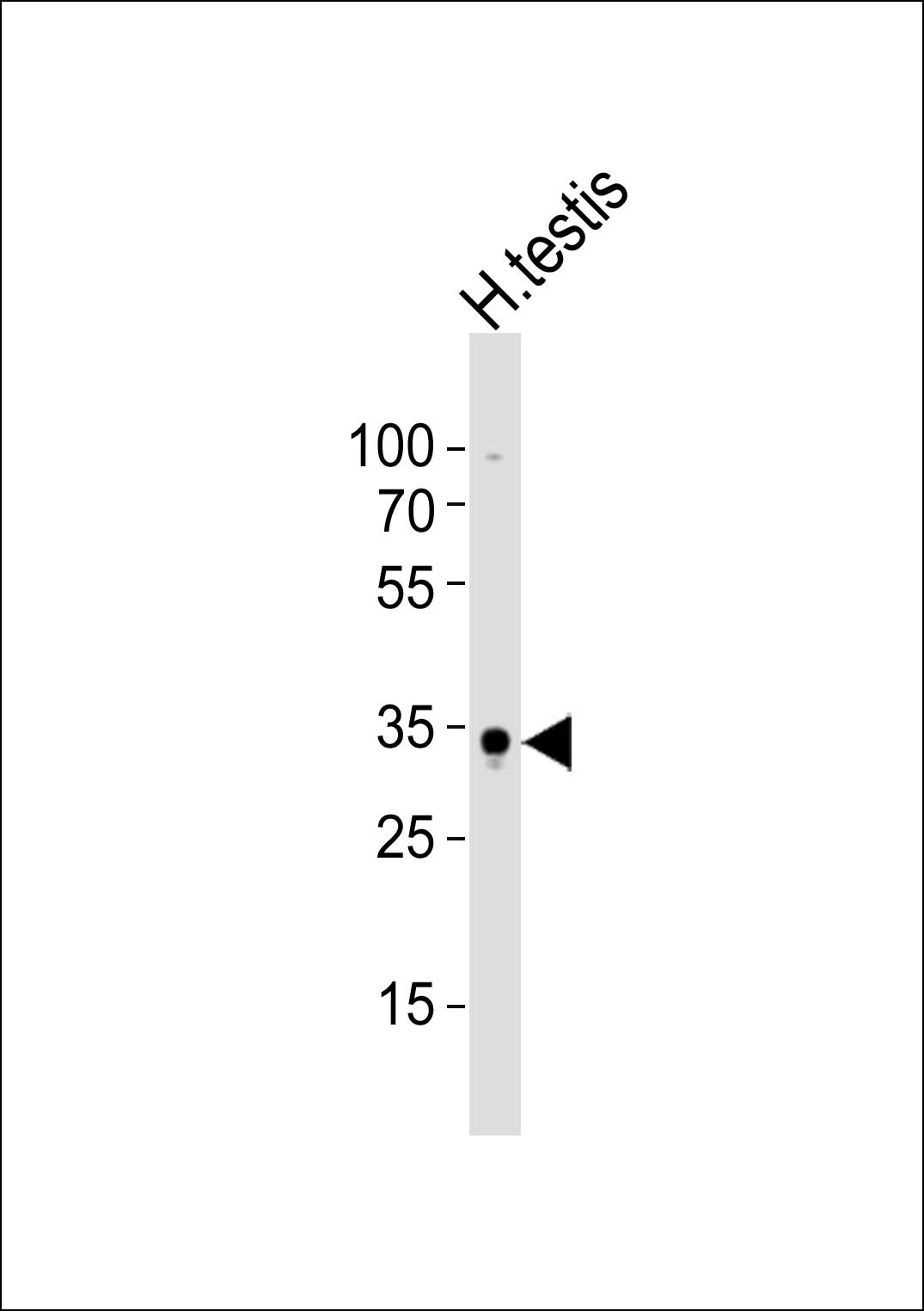
(Mouse) Sox15 Antibody (C-term)
Purified Rabbit Polyclonal Antibody (Pab)
- SPECIFICATION
- CITATIONS
- PROTOCOLS
- BACKGROUND

Application
| WB, E |
|---|---|
| Primary Accession | P43267 |
| Reactivity | Human |
| Host | Rabbit |
| Clonality | Polyclonal |
| Isotype | Rabbit IgG |
| Calculated MW | 25311 Da |
| Antigen Region | 152-165 aa |
| Gene ID | 20670 |
|---|---|
| Other Names | Protein SOX-15, Sox15, Sox-15 |
| Target/Specificity | This (Mouse) Sox15 antibody is generated from a rabbit immunized with a KLH conjugated synthetic peptide between 152-165 amino acids from the C-terminal region of human (Mouse) Sox15. |
| Dilution | WB~~1:1000 E~~Use at an assay dependent concentration. |
| Format | Purified polyclonal antibody supplied in PBS with 0.09% (W/V) sodium azide. This antibody is purified through a protein A column, followed by peptide affinity purification. |
| Storage | Maintain refrigerated at 2-8°C for up to 2 weeks. For long term storage store at -20°C in small aliquots to prevent freeze-thaw cycles. |
| Precautions | (Mouse) Sox15 Antibody (C-term) is for research use only and not for use in diagnostic or therapeutic procedures. |
| Name | Sox15 {ECO:0000312|MGI:MGI:98363} |
|---|---|
| Function | Transcription factor that binds to DNA at the 5'-AACAATG-3' consensus sequence (PubMed:10821863, PubMed:15863505, PubMed:16759287, PubMed:17363903). Acts as a transcriptional activator and repressor (PubMed:10821863, PubMed:15863505, PubMed:16759287). Binds synergistically with POU5F1 (OCT3/4) to gene promoters (PubMed:15863505). Binds to the FOXK1 promoter and recruits FHL3, resulting in transcriptional activation of FOXK1 which leads to myoblast proliferation (PubMed:17363903). Acts as an inhibitor of myoblast differentiation via transcriptional repression which leads to down-regulation of the muscle-specific genes MYOD and MYOG (PubMed:10821863). Involved in trophoblast giant cell differentiation via enhancement of HAND1 transcriptional activity (PubMed:16759287). Regulates transcription of HRC via binding to its proximal enhancer region (PubMed:15863505). Involved in skeletal muscle regeneration (PubMed:15367664, PubMed:17363903). Also plays a role in the development of myogenic precursor cells (PubMed:15367664). |
| Cellular Location | Nucleus {ECO:0000255|PROSITE-ProRule:PRU00267, ECO:0000269|PubMed:10821863, ECO:0000269|PubMed:15367664, ECO:0000269|PubMed:17363903} |
| Tissue Location | Expressed in myoblasts (at protein level) (PubMed:15367664). Expressed in embryonic stem cells (at protein level) (PubMed:15367664, PubMed:15863505). Expressed in myogenic progenitor cells (at protein level) (PubMed:17363903). Expressed in the ovary (PubMed:15367664). Expressed in kidney, liver, skeletal muscle, and testes (PubMed:10821863, PubMed:15367664). Expressed in lung and skin (PubMed:15863505). Expressed in the brain, heart, diaphragm, and intestines (PubMed:10821863). Expressed in the conceptus tissues of the placenta (PubMed:16759287). |

Thousands of laboratories across the world have published research that depended on the performance of antibodies from Abcepta to advance their research. Check out links to articles that cite our products in major peer-reviewed journals, organized by research category.
info@abcepta.com, and receive a free "I Love Antibodies" mug.

Provided below are standard protocols that you may find useful for product applications.
References
Miyashita A.,et al.Gene 237:53-60(1999).
Beranger F.,et al.J. Biol. Chem. 275:16103-16109(2000).
Liu Y.,et al.Submitted (FEB-2000) to the EMBL/GenBank/DDBJ databases.
Stock D.W.,et al.Genomics 37:234-237(1996).
van de Wetering M.,et al.Nucleic Acids Res. 21:1669-1669(1993).
If you have used an Abcepta product and would like to share how it has performed, please click on the "Submit Review" button and provide the requested information. Our staff will examine and post your review and contact you if needed.
If you have any additional inquiries please email technical services at tech@abcepta.com.
Ordering Information
Other Products
Shipping Information
