


 Foundational characteristics of cancer include proliferation, angiogenesis, migration, evasion of apoptosis, and cellular immortality. Find key markers for these cellular processes and antibodies to detect them.
 The SUMOplot™ Analysis Program predicts and scores sumoylation sites in your protein. SUMOylation is a post-translational modification involved in various cellular processes, such as nuclear-cytosolic transport, transcriptional regulation, apoptosis, protein stability, response to stress, and progression through the cell cycle.
 The Autophagy Receptor Motif Plotter predicts and scores autophagy receptor binding sites in your protein. Identifying proteins connected to this pathway is critical to understanding the role of autophagy in physiological as well as pathological processes such as development, differentiation, neurodegenerative diseases, stress, infection, and cancer.
SMURF1 Antibody (N-term)
Purified Rabbit Polyclonal Antibody (Pab)
- SPECIFICATION
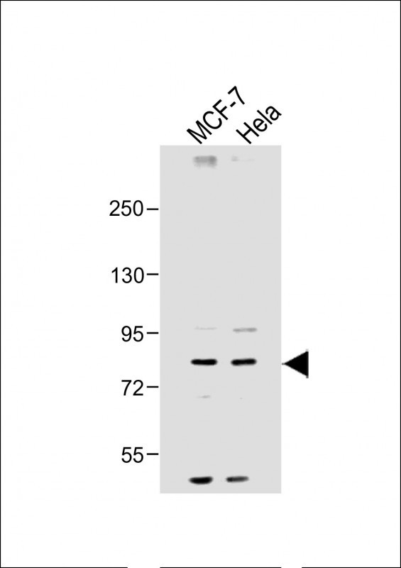
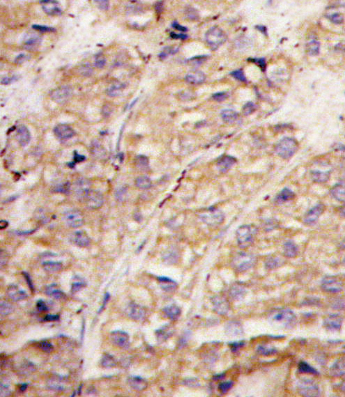
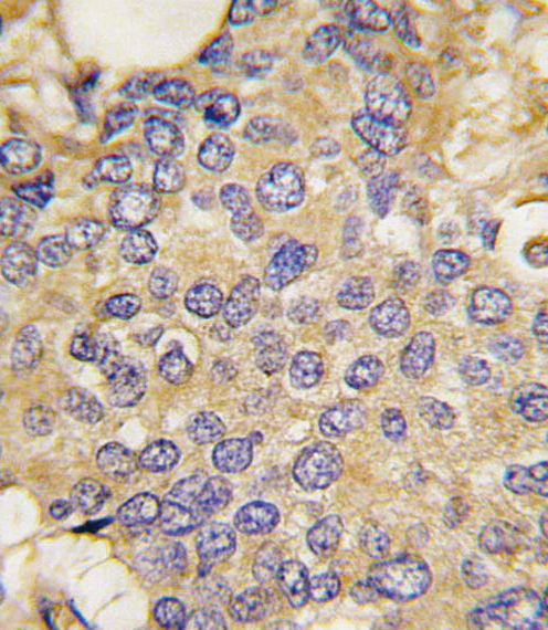
- CITATIONS: 4
- PROTOCOLS
- BACKGROUND

Application
| IHC-P, WB, E |
|---|---|
| Primary Accession | Q9HCE7 |
| Other Accession | Q2TAS2, A2A5Z6, Q9HAU4, A9JRZ0, Q9PUN2, Q9CUN6 |
| Reactivity | Human, Mouse |
| Predicted | Xenopus, Zebrafish |
| Host | Rabbit |
| Clonality | Polyclonal |
| Isotype | Rabbit IgG |
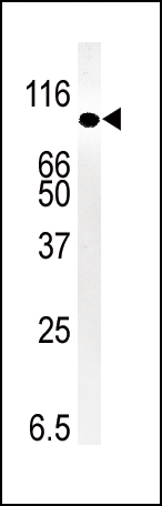
| Calculated MW | 86114 Da |
| Antigen Region | 66-96 aa |
| Gene ID | 57154 |
|---|---|
| Other Names | E3 ubiquitin-protein ligase SMURF1, hSMURF1, 632-, SMAD ubiquitination regulatory factor 1, SMAD-specific E3 ubiquitin-protein ligase 1, SMURF1, KIAA1625 |
| Target/Specificity | This SMURF1 antibody is generated from rabbits immunized with a KLH conjugated synthetic peptide between 66-96 amino acids from the N-terminal region of human SMURF1. |
| Dilution | IHC-P~~1:10~50 WB~~1:2000 E~~Use at an assay dependent concentration. |
| Format | Purified polyclonal antibody supplied in PBS with 0.09% (W/V) sodium azide. This antibody is prepared by Saturated Ammonium Sulfate (SAS) precipitation followed by dialysis against PBS. |
| Storage | Maintain refrigerated at 2-8°C for up to 2 weeks. For long term storage store at -20°C in small aliquots to prevent freeze-thaw cycles. |
| Precautions | SMURF1 Antibody (N-term) is for research use only and not for use in diagnostic or therapeutic procedures. |
| Name | SMURF1 |
|---|---|
| Synonyms | KIAA1625 |
| Function | E3 ubiquitin-protein ligase that acts as a negative regulator of BMP signaling pathway. Mediates ubiquitination and degradation of SMAD1 and SMAD5, 2 receptor-regulated SMADs specific for the BMP pathway. Promotes ubiquitination and subsequent proteasomal degradation of TRAF family members and RHOA. Promotes ubiquitination and subsequent proteasomal degradation of MAVS (PubMed:23087404). Acts as an antagonist of TGF-beta signaling by ubiquitinating TGFBR1 and targeting it for degradation (PubMed:21791611). Plays a role in dendrite formation by melanocytes (PubMed:23999003). |
| Cellular Location | Cytoplasm. Cell membrane; Peripheral membrane protein; Cytoplasmic side |
| Tissue Location | Expressed in melanocytes (PubMed:23999003). |

Provided below are standard protocols that you may find useful for product applications.
Background
Members of the transforming growth factor-beta (TGFB) family signal through type I and type II serine/threonine-kinase receptors, which in turn activate the SMAD signaling pathway. Bone morphogenetic protein (BMP) receptors target SMAD1, SMAD5, and SMAD8, whereas receptors for activin and TGFB (e.g., ACVR1 and TGFBR1, respectively) target SMAD2 and SMAD3. Phosphorylation of these receptor-regulated SMADs induces their association with the common-partner SMAD, SMAD4. Smurf1, a HECT domain E3 ubiquitin ligase, regulates cell polarity and protrusive activity and is required to maintain the transformed morphology and motility of a tumor cell. Atypical protein kinase C-zeta (PKC2), an effector of the Cdc42/Rac1-PAR6 polarity complex, recruits Smurf1 to cellular protrusions, where it controlled the local level of RhoA. Smurf1 thus links the polarity complex to degradation of RhoA in lamellipodia and filopodia to prevent RhoA signaling during dynamic membrane movements.
References
Tajima, Y., et al., J. Biol. Chem. 278(12):10716-10721 (2003). Suzuki, C., et al., J. Biol. Chem. 277(42):39919-39925 (2002). Ebisawa, T., et al., J. Biol. Chem. 276(16):12477-12480 (2001). Zhu, H., et al., Nature 400(6745):687-693 (1999). Lambris, J., et al., J. Immunol. Methods 27(1):55-59 (1979).
If you have used an Abcepta product and would like to share how it has performed, please click on the "Submit Review" button and provide the requested information. Our staff will examine and post your review and contact you if needed.
If you have any additional inquiries please email technical services at tech@abcepta.com.
Ordering Information
Other Products
Shipping Information
