


 Foundational characteristics of cancer include proliferation, angiogenesis, migration, evasion of apoptosis, and cellular immortality. Find key markers for these cellular processes and antibodies to detect them.
 The SUMOplot™ Analysis Program predicts and scores sumoylation sites in your protein. SUMOylation is a post-translational modification involved in various cellular processes, such as nuclear-cytosolic transport, transcriptional regulation, apoptosis, protein stability, response to stress, and progression through the cell cycle.
 The Autophagy Receptor Motif Plotter predicts and scores autophagy receptor binding sites in your protein. Identifying proteins connected to this pathway is critical to understanding the role of autophagy in physiological as well as pathological processes such as development, differentiation, neurodegenerative diseases, stress, infection, and cancer.
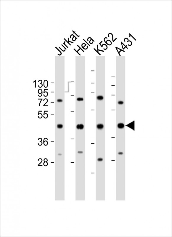
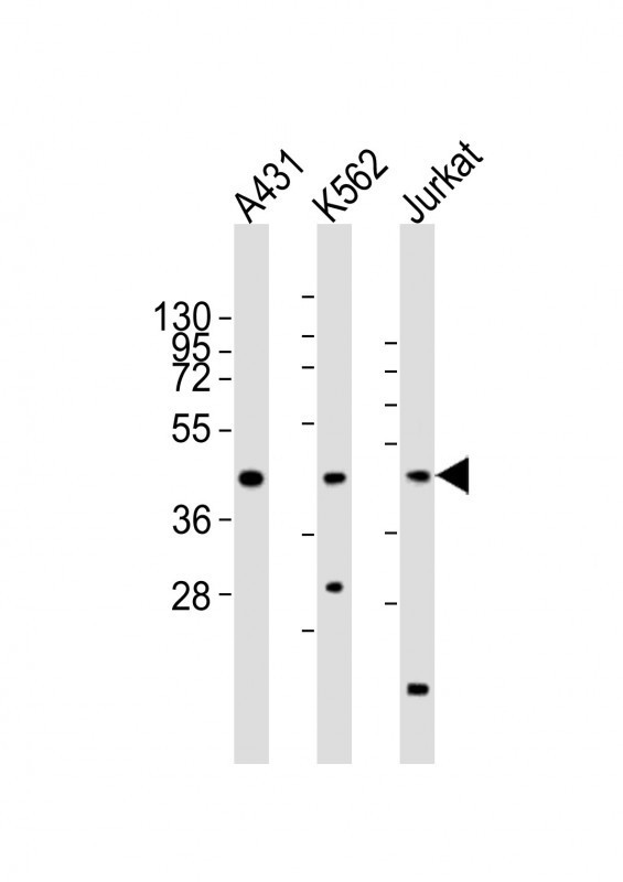
RBMX Antibody (Center)
Purified Rabbit Polyclonal Antibody (Pab)
- SPECIFICATION
- CITATIONS: 1
- PROTOCOLS
- BACKGROUND

Application
| WB, E |
|---|---|
| Primary Accession | P38159 |
| Reactivity | Human |
| Host | Rabbit |
| Clonality | polyclonal |
| Isotype | Rabbit IgG |
| Calculated MW | 42332 Da |
| Gene ID | 27316 |
|---|---|
| Other Names | RNA-binding motif protein, X chromosome, Glycoprotein p43, Heterogeneous nuclear ribonucleoprotein G, hnRNP G, RNA-binding motif protein, X chromosome, N-terminally processed, RBMX, HNRPG, RBMXP1 |
| Target/Specificity | This RBMX antibody is generated from a rabbit immunized with a KLH conjugated synthetic peptide between 222-252 amino acids from the Central region of human RBMX. |
| Dilution | WB~~1:2000 E~~Use at an assay dependent concentration. |
| Format | Purified polyclonal antibody supplied in PBS with 0.09% (W/V) sodium azide. This antibody is purified through a protein A column, followed by peptide affinity purification. |
| Storage | Maintain refrigerated at 2-8°C for up to 2 weeks. For long term storage store at -20°C in small aliquots to prevent freeze-thaw cycles. |
| Precautions | RBMX Antibody (Center) is for research use only and not for use in diagnostic or therapeutic procedures. |
| Name | RBMX |
|---|---|
| Synonyms | HNRPG, RBMXP1 |
| Function | RNA-binding protein that plays several role in the regulation of pre- and post-transcriptional processes. Implicated in tissue- specific regulation of gene transcription and alternative splicing of several pre-mRNAs. Binds to and stimulates transcription from the tumor suppressor TXNIP gene promoter; may thus be involved in tumor suppression. When associated with SAFB, binds to and stimulates transcription from the SREBF1 promoter. Associates with nascent mRNAs transcribed by RNA polymerase II. Component of the supraspliceosome complex that regulates pre-mRNA alternative splice site selection. Can either activate or suppress exon inclusion; acts additively with TRA2B to promote exon 7 inclusion of the survival motor neuron SMN2. Represses the splicing of MAPT/Tau exon 10. Binds preferentially to single-stranded 5'-CC[A/C]-rich RNA sequence motifs localized in a single-stranded conformation; probably binds RNA as a homodimer. Binds non-specifically to pre-mRNAs. Also plays a role in the cytoplasmic TNFR1 trafficking pathways; promotes both the IL-1-beta-mediated inducible proteolytic cleavage of TNFR1 ectodomains and the release of TNFR1 exosome-like vesicles to the extracellular compartment. |
| Cellular Location | Nucleus Note=Component of ribonucleosomes. Localizes in numerous small granules in the nucleus |
| Tissue Location | Expressed strongly in oral keratinocytes, but only weakly detected in oral squamous cell carcinomas (at protein level) |

Provided below are standard protocols that you may find useful for product applications.
Background
RNA-binding protein that plays several role in the regulation of pre- and post-transcriptional processes. Implicated in tissue-specific regulation of gene transcription and alternative splicing of several pre-mRNAs. Binds to and stimulates transcription from the tumor suppressor TXNIP gene promoter; may thus be involved in tumor suppression. When associated with SAFB, binds to and stimulates transcription from the SREBF1 promoter. Associates with nascent mRNAs transcribed by RNA polymerase II. Component of the supraspliceosome complex that regulates pre-mRNA alternative splice site selection. Can either activate or suppress exon inclusion; acts additively with TRA2B to promote exon 7 inclusion of the survival motor neuron SMN2. Represses the splicing of MAPT/Tau exon 10. Binds preferentially to single- stranded 5'-CC[A/C]-rich RNA sequence motifs localized in a single-stranded conformation; probably binds RNA as a homodimer. Binds non-specifically to pre-mRNAs. Plays also a role in the cytoplasmic TNFR1 trafficking pathways; promotes both the IL-1- beta-mediated inducible proteolytic cleavage of TNFR1 ectodomains and the release of TNFR1 exosome-like vesicles to the extracellular compartment.
References
Soulard M.,et al.Nucleic Acids Res. 21:4210-4217(1993).
Venables J.P.,et al.Submitted (JUN-1998) to the EMBL/GenBank/DDBJ databases.
Lingenfelter P.A.,et al.Mamm. Genome 12:538-545(2001).
Lin T.-Y.,et al.Submitted (NOV-2003) to the EMBL/GenBank/DDBJ databases.
Ota T.,et al.Nat. Genet. 36:40-45(2004).
If you have used an Abcepta product and would like to share how it has performed, please click on the "Submit Review" button and provide the requested information. Our staff will examine and post your review and contact you if needed.
If you have any additional inquiries please email technical services at tech@abcepta.com.
Ordering Information
Other Products
Shipping Information
