


 Foundational characteristics of cancer include proliferation, angiogenesis, migration, evasion of apoptosis, and cellular immortality. Find key markers for these cellular processes and antibodies to detect them.
 The SUMOplot™ Analysis Program predicts and scores sumoylation sites in your protein. SUMOylation is a post-translational modification involved in various cellular processes, such as nuclear-cytosolic transport, transcriptional regulation, apoptosis, protein stability, response to stress, and progression through the cell cycle.
 The Autophagy Receptor Motif Plotter predicts and scores autophagy receptor binding sites in your protein. Identifying proteins connected to this pathway is critical to understanding the role of autophagy in physiological as well as pathological processes such as development, differentiation, neurodegenerative diseases, stress, infection, and cancer.
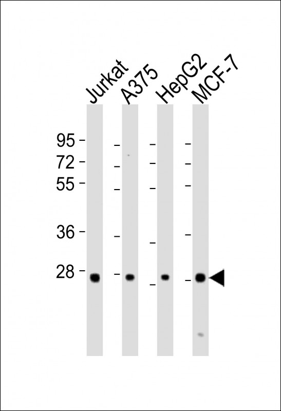
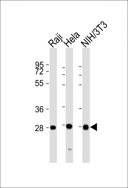
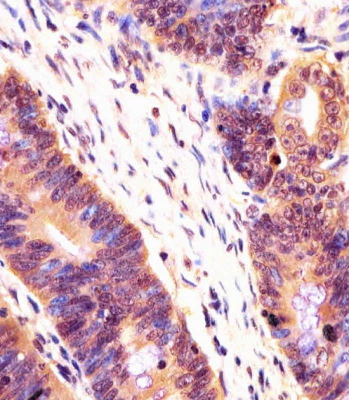
PSMB7 Antibody (Center)
Purified Rabbit Polyclonal Antibody (Pab)
- SPECIFICATION
- CITATIONS: 1
- PROTOCOLS
- BACKGROUND

Application
| WB, IHC-P, E |
|---|---|
| Primary Accession | Q99436 |
| Reactivity | Human, Mouse |
| Host | Rabbit |
| Clonality | polyclonal |
| Isotype | Rabbit IgG |
| Calculated MW | 29965 Da |
| Gene ID | 5695 |
|---|---|
| Other Names | Proteasome subunit beta type-7, Macropain chain Z, Multicatalytic endopeptidase complex chain Z, Proteasome subunit Z, PSMB7, Z |
| Target/Specificity | This PSMB7 antibody is generated from a rabbit immunized with a KLH conjugated synthetic peptide between 180-211 amino acids from the Central region of human PSMB7. |
| Dilution | WB~~1:2000 IHC-P~~1:25 E~~Use at an assay dependent concentration. |
| Format | Purified polyclonal antibody supplied in PBS with 0.09% (W/V) sodium azide. This antibody is purified through a protein A column, followed by peptide affinity purification. |
| Storage | Maintain refrigerated at 2-8°C for up to 2 weeks. For long term storage store at -20°C in small aliquots to prevent freeze-thaw cycles. |
| Precautions | PSMB7 Antibody (Center) is for research use only and not for use in diagnostic or therapeutic procedures. |
| Name | PSMB7 (HGNC:9544) |
|---|---|
| Synonyms | Z |
| Function | Component of the 20S core proteasome complex involved in the proteolytic degradation of most intracellular proteins. This complex plays numerous essential roles within the cell by associating with different regulatory particles. Associated with two 19S regulatory particles, forms the 26S proteasome and thus participates in the ATP- dependent degradation of ubiquitinated proteins. The 26S proteasome plays a key role in the maintenance of protein homeostasis by removing misfolded or damaged proteins that could impair cellular functions, and by removing proteins whose functions are no longer required. Associated with the PA200 or PA28, the 20S proteasome mediates ubiquitin- independent protein degradation. This type of proteolysis is required in several pathways including spermatogenesis (20S-PA200 complex) or generation of a subset of MHC class I-presented antigenic peptides (20S-PA28 complex). Within the 20S core complex, PSMB7 displays a trypsin-like activity. |
| Cellular Location | Cytoplasm. Nucleus. Note=Translocated from the cytoplasm into the nucleus following interaction with AKIRIN2, which bridges the proteasome with the nuclear import receptor IPO9 |
| Tissue Location | Expressed at a low level in colonic mucosa. Up- regulated in colorectal cancer tissues. |

Provided below are standard protocols that you may find useful for product applications.
Background
The proteasome is a multicatalytic proteinase complex which is characterized by its ability to cleave peptides with Arg, Phe, Tyr, Leu, and Glu adjacent to the leaving group at neutral or slightly basic pH. The proteasome has an ATP-dependent proteolytic activity. This unit is responsible of the trypsin-like activity.
References
Hisamatsu H.,et al.J. Exp. Med. 183:1807-1816(1996).
Ota T.,et al.Nat. Genet. 36:40-45(2004).
Humphray S.J.,et al.Nature 429:369-374(2004).
Mural R.J.,et al.Submitted (JUL-2005) to the EMBL/GenBank/DDBJ databases.
Lubec G.,et al.Submitted (DEC-2008) to UniProtKB.
If you have used an Abcepta product and would like to share how it has performed, please click on the "Submit Review" button and provide the requested information. Our staff will examine and post your review and contact you if needed.
If you have any additional inquiries please email technical services at tech@abcepta.com.
Ordering Information
Other Products
Shipping Information
