


 Foundational characteristics of cancer include proliferation, angiogenesis, migration, evasion of apoptosis, and cellular immortality. Find key markers for these cellular processes and antibodies to detect them.
 The SUMOplot™ Analysis Program predicts and scores sumoylation sites in your protein. SUMOylation is a post-translational modification involved in various cellular processes, such as nuclear-cytosolic transport, transcriptional regulation, apoptosis, protein stability, response to stress, and progression through the cell cycle.
 The Autophagy Receptor Motif Plotter predicts and scores autophagy receptor binding sites in your protein. Identifying proteins connected to this pathway is critical to understanding the role of autophagy in physiological as well as pathological processes such as development, differentiation, neurodegenerative diseases, stress, infection, and cancer.
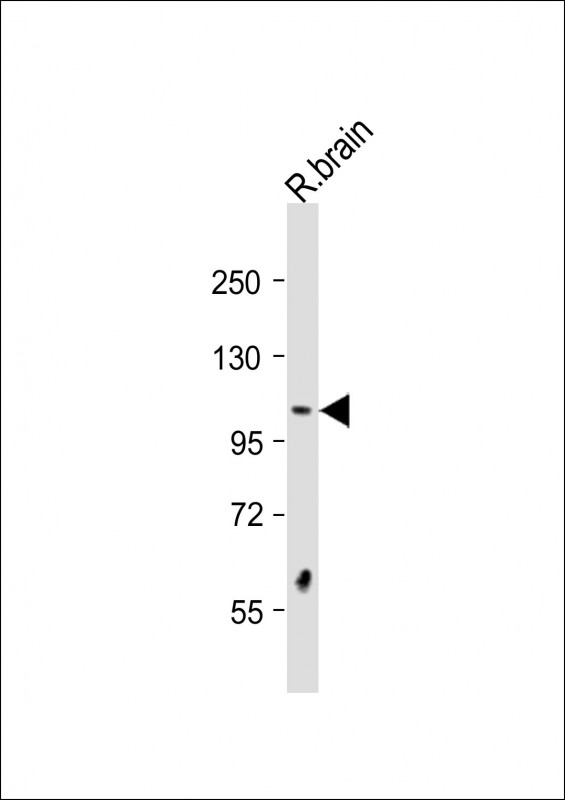
DSTYK Antibody (N-Term)
Purified Rabbit Polyclonal Antibody (Pab)
- SPECIFICATION
- CITATIONS
- PROTOCOLS
- BACKGROUND

Application
| WB, E |
|---|---|
| Primary Accession | Q6XUX3 |
| Reactivity | Rat |
| Host | Rabbit |
| Clonality | polyclonal |
| Isotype | Rabbit IgG |
| Calculated MW | 105206 Da |
| Gene ID | 25778 |
|---|---|
| Other Names | Dual serine/threonine and tyrosine protein kinase, Dusty protein kinase, Dusty PK, RIP-homologous kinase, Receptor-interacting serine/threonine-protein kinase 5, Sugen kinase 496, SgK496, DSTYK, KIAA0472, RIP5, RIPK5, SGK496 |
| Target/Specificity | This DSTYK antibody is generated from a rabbit immunized with a KLH conjugated synthetic peptide between 256-290 amino acids from human DSTYK. |
| Dilution | WB~~1:2000 E~~Use at an assay dependent concentration. |
| Format | Purified polyclonal antibody supplied in PBS with 0.09% (W/V) sodium azide. This antibody is purified through a protein A column, followed by peptide affinity purification. |
| Storage | Maintain refrigerated at 2-8°C for up to 2 weeks. For long term storage store at -20°C in small aliquots to prevent freeze-thaw cycles. |
| Precautions | DSTYK Antibody (N-Term) is for research use only and not for use in diagnostic or therapeutic procedures. |
| Name | DSTYK |
|---|---|
| Synonyms | KIAA0472, RIP5, RIPK5, SGK496 |
| Function | Acts as a positive regulator of ERK phosphorylation downstream of fibroblast growth factor-receptor activation (PubMed:23862974, PubMed:28157540). Involved in the regulation of both caspase-dependent apoptosis and caspase-independent cell death (PubMed:15178406). In the skin, it plays a predominant role in suppressing caspase-dependent apoptosis in response to UV stress in a range of dermal cell types (PubMed:28157540). |
| Cellular Location | Cytoplasm. Cell membrane {ECO:0000250|UniProtKB:Q6XUX1}. Apical cell membrane. Basolateral cell membrane. Cell junction {ECO:0000250|UniProtKB:Q6XUX1}. Note=Detected at apical cell-cell junctions. Colocalized with FGF receptors to the cell membrane (By similarity). Detected in basolateral and apical membranes of all tubular epithelia. {ECO:0000250|UniProtKB:Q6XUX1, ECO:0000269|PubMed:23862974} |
| Tissue Location | Predominantly expressed in skeletal muscle and testis. Expressed in basolateral and apical membranes of all tubular epithelia. Expressed in thin ascending limb of the loop of Henle and the distal convoluted tubule. Expressed in all layers of transitional ureteric epithelium and in the ureteric smooth-muscle cells. Weakly expressed in heart, brain, placenta, kidney, pancreas, spleen, thymus, prostate, uterus, small intestine, white blood cells, stomach, spinal cord and adrenal gland. Is widely distributed in the CNS. Also detected in several tumor cell lines. Expressed in the skin (PubMed:28157540) |

Thousands of laboratories across the world have published research that depended on the performance of antibodies from Abcepta to advance their research. Check out links to articles that cite our products in major peer-reviewed journals, organized by research category.
info@abcepta.com, and receive a free "I Love Antibodies" mug.

Provided below are standard protocols that you may find useful for product applications.
Background
Acts as a positive regulator of ERK phosphorylation downstream of fibroblast growth factor-receptor activation. May induce both caspase-dependent apoptosis and caspase-independent cell death.
References
Peng J.,et al.Biochim. Biophys. Acta 1759:562-572(2006).
Zhao Z.,et al.Submitted (MAY-1998) to the EMBL/GenBank/DDBJ databases.
Gregory S.G.,et al.Nature 441:315-321(2006).
Seki N.,et al.DNA Res. 4:345-349(1997).
Zha J.,et al.Biochem. Biophys. Res. Commun. 319:298-303(2004).
If you have used an Abcepta product and would like to share how it has performed, please click on the "Submit Review" button and provide the requested information. Our staff will examine and post your review and contact you if needed.
If you have any additional inquiries please email technical services at tech@abcepta.com.
Ordering Information
Other Products
Shipping Information
