


 Foundational characteristics of cancer include proliferation, angiogenesis, migration, evasion of apoptosis, and cellular immortality. Find key markers for these cellular processes and antibodies to detect them.
 The SUMOplot™ Analysis Program predicts and scores sumoylation sites in your protein. SUMOylation is a post-translational modification involved in various cellular processes, such as nuclear-cytosolic transport, transcriptional regulation, apoptosis, protein stability, response to stress, and progression through the cell cycle.
 The Autophagy Receptor Motif Plotter predicts and scores autophagy receptor binding sites in your protein. Identifying proteins connected to this pathway is critical to understanding the role of autophagy in physiological as well as pathological processes such as development, differentiation, neurodegenerative diseases, stress, infection, and cancer.
BAP1 Antibody (N-term)
Purified Rabbit Polyclonal Antibody (Pab)

- SPECIFICATION
- CITATIONS: 1
- PROTOCOLS
- BACKGROUND

Application
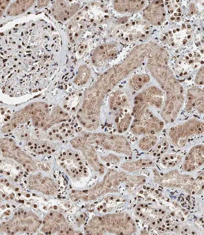
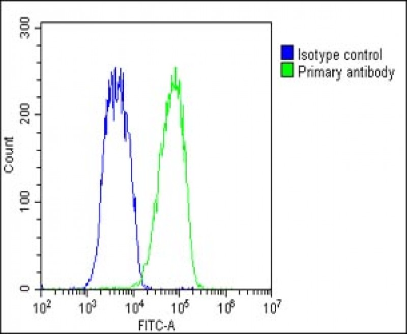
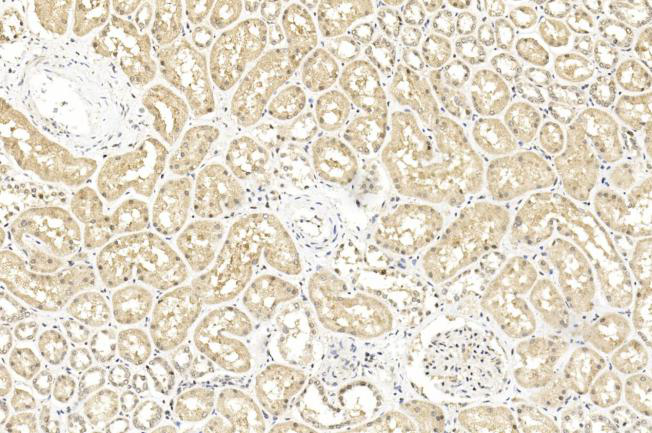
| WB, IHC-P, IF, FC, E |
|---|---|
| Primary Accession | Q92560 |
| Other Accession | D3ZHS6, Q99PU7, A1L2G3, A2VDM8 |
| Reactivity | Human, Mouse, Rat |
| Predicted | Bovine, Zebrafish |
| Host | Rabbit |
| Clonality | Polyclonal |
| Isotype | Rabbit IgG |
| Calculated MW | 80362 Da |
| Antigen Region | 36-66 aa |
| Gene ID | 8314 |
|---|---|
| Other Names | Ubiquitin carboxyl-terminal hydrolase BAP1, BRCA1-associated protein 1, Cerebral protein 6, BAP1, KIAA0272 |
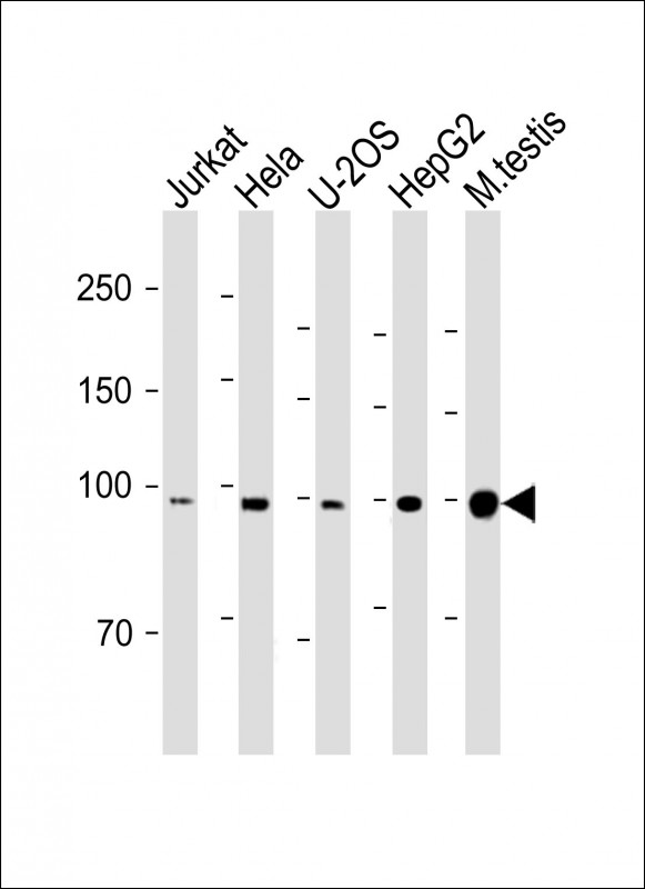
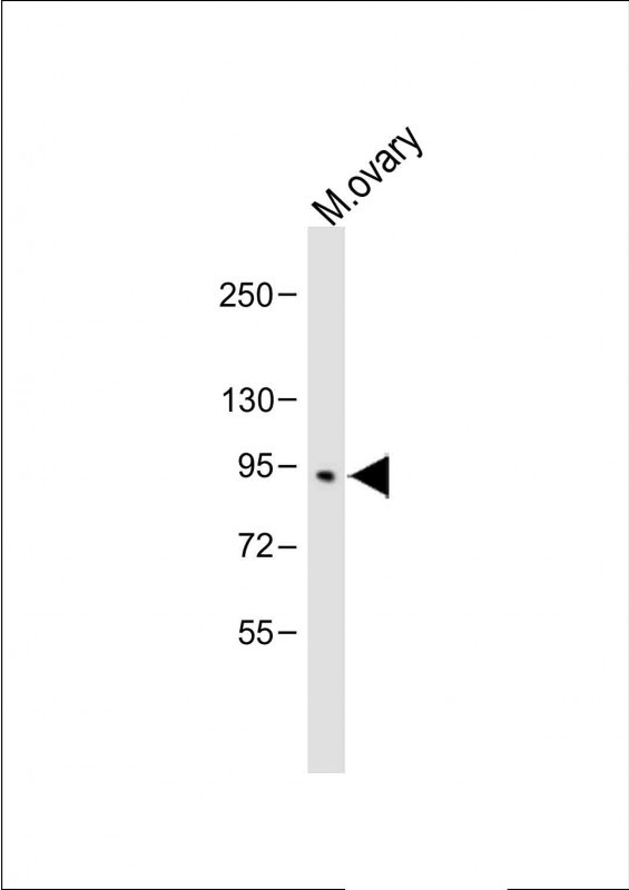
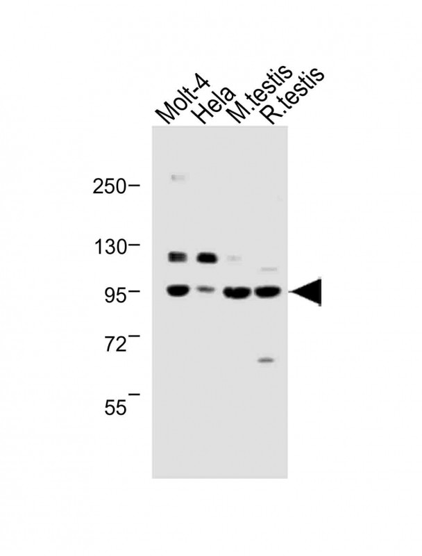
| Target/Specificity | This BAP1 antibody is generated from rabbits immunized with a KLH conjugated synthetic peptide between 36-66 amino acids of human BAP1. |
| Dilution | WB~~1:1000 IHC-P~~1:100 IF~~1:25 FC~~1:25 E~~Use at an assay dependent concentration. |
| Format | Purified polyclonal antibody supplied in PBS with 0.09% (W/V) sodium azide. This antibody is purified through a protein A column, followed by peptide affinity purification. |
| Storage | Maintain refrigerated at 2-8°C for up to 2 weeks. For long term storage store at -20°C in small aliquots to prevent freeze-thaw cycles. |
| Precautions | BAP1 Antibody (N-term) is for research use only and not for use in diagnostic or therapeutic procedures. |
| Name | BAP1 {ECO:0000303|PubMed:9528852, ECO:0000312|HGNC:HGNC:950} |
|---|---|
| Function | Deubiquitinating enzyme that plays a key role in chromatin by mediating deubiquitination of histone H2A and HCFC1 (PubMed:12485996, PubMed:18757409, PubMed:20436459, PubMed:25451922, PubMed:35051358). Catalytic component of the polycomb repressive deubiquitinase (PR-DUB) complex, a complex that specifically mediates deubiquitination of histone H2A monoubiquitinated at 'Lys-120' (H2AK119ub1) (PubMed:20436459, PubMed:25451922, PubMed:30664650, PubMed:35051358). Does not deubiquitinate monoubiquitinated histone H2B (PubMed:20436459, PubMed:30664650). The PR-DUB complex is an epigenetic regulator of gene expression and acts as a transcriptional coactivator, affecting genes involved in development, cell communication, signaling, cell proliferation and cell viability (PubMed:20805357, PubMed:30664650, PubMed:36180891). Antagonizes PRC1 mediated H2AK119ub1 monoubiquitination (PubMed:30664650). As part of the PR-DUB complex, associates with chromatin enriched in histone marks H3K4me1, H3K4me3, and H3K27Ac, but not in H3K27me3 (PubMed:36180891). Recruited to specific gene-regulatory regions by YY1 (PubMed:20805357). Acts as a regulator of cell growth by mediating deubiquitination of HCFC1 N- terminal and C-terminal chains, with some specificity toward 'Lys-48'- linked polyubiquitin chains compared to 'Lys-63'-linked polyubiquitin chains (PubMed:19188440, PubMed:19815555). Deubiquitination of HCFC1 does not lead to increase stability of HCFC1 (PubMed:19188440, PubMed:19815555). Interferes with the BRCA1 and BARD1 heterodimer activity by inhibiting their ability to mediate ubiquitination and autoubiquitination (PubMed:19117993). It however does not mediate deubiquitination of BRCA1 and BARD1 (PubMed:19117993). Able to mediate autodeubiquitination via intramolecular interactions to counteract monoubiquitination at the nuclear localization signal (NLS), thereby protecting it from cytoplasmic sequestration (PubMed:24703950). Negatively regulates epithelial-mesenchymal transition (EMT) of trophoblast stem cells during placental development by regulating genes involved in epithelial cell integrity, cell adhesion and cytoskeletal organization (PubMed:34170818). |
| Cellular Location | Cytoplasm. Nucleus. Chromosome. Note=Mainly nuclear (PubMed:24703950, PubMed:30664650). Binds to chromatin (PubMed:30664650). Localizes to the cytoplasm when monoubiquitinated by the E2/E3 hybrid ubiquitin- protein ligase UBE2O (PubMed:24703950). Recruitment to chromatin is dependent on ASXL1/2/3 and recruitment to specific genes on FOXK1/2 (By similarity). Nuclear localization is redundantly mediated by the importin and transportin systems; TNPO1/transportin-1 is the major mediator of nuclear localization (PubMed:35446349) {ECO:0000250|UniProtKB:Q99PU7, ECO:0000269|PubMed:24703950, ECO:0000269|PubMed:30664650, ECO:0000269|PubMed:35446349} |
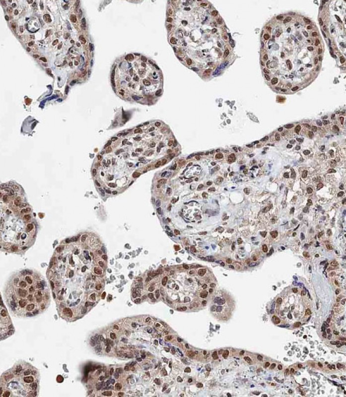
| Tissue Location | Highly expressed in testis, placenta and ovary (PubMed:9528852). Expressed in breast (PubMed:9528852). levels in the placenta increase over the course of pregnancy (PubMed:34170818) |

Provided below are standard protocols that you may find useful for product applications.
Background
'BRCA1-associated protein-1,' or BAP1 interacts with the RING finger domain of BRCA1. The N-terminal 240 amino acids of the predicted 729-amino acid human protein show homology to ubiquitin C-terminal hydrolases (UCHs), thiol proteases that catalyze proteolytic processing of ubiquitin. In addition, BAP1 contains an acidic region, a highly charged C-terminal region, and 2 putative nuclear localization signals.. BAP1 and BRCA1 associate in vivo and have overlapping subnuclear localization patterns.1 BAP1 enhances BRCA1-mediated inhibition of breast cancer cell growth. Northern blot analysis indicates that BAP1 is expressed as a 4-kb mRNA in all human tissues tested, with A 4.8-kb transcript expressed exclusively in testis. Northern blot analysis and in situ hybridization reveal that BAP1 and BRCA1 are coexpressed during murine breast development and remodeling. The BAP1 gene has been mapped to 3p21.3, a region of loss of heterozygosity for breast cancer as well as frequently deleted in lung carcinomas.1 Intragenic homozygous rearrangements and deletions of BAP1 appear in lung carcinoma cell lines. It has been postulated that BAP1 is a tumor suppressor gene that functions in the BRCA1 growth control pathway.1
If you have used an Abcepta product and would like to share how it has performed, please click on the "Submit Review" button and provide the requested information. Our staff will examine and post your review and contact you if needed.
If you have any additional inquiries please email technical services at tech@abcepta.com.
Ordering Information
Other Products
Shipping Information
