


 Foundational characteristics of cancer include proliferation, angiogenesis, migration, evasion of apoptosis, and cellular immortality. Find key markers for these cellular processes and antibodies to detect them.
 The SUMOplot™ Analysis Program predicts and scores sumoylation sites in your protein. SUMOylation is a post-translational modification involved in various cellular processes, such as nuclear-cytosolic transport, transcriptional regulation, apoptosis, protein stability, response to stress, and progression through the cell cycle.
 The Autophagy Receptor Motif Plotter predicts and scores autophagy receptor binding sites in your protein. Identifying proteins connected to this pathway is critical to understanding the role of autophagy in physiological as well as pathological processes such as development, differentiation, neurodegenerative diseases, stress, infection, and cancer.
BATF3 Antibody (Center)
Purified Rabbit Polyclonal Antibody (Pab)
- SPECIFICATION
- CITATIONS
- PROTOCOLS
- BACKGROUND

Application
| WB, E |
|---|---|
| Primary Accession | Q9NR55 |
| Reactivity | Human |
| Host | Rabbit |
| Clonality | polyclonal |
| Isotype | Rabbit IgG |
| Calculated MW | 14468 Da |
| Gene ID | 55509 |
|---|---|
| Other Names | Basic leucine zipper transcriptional factor ATF-like 3, B-ATF-3, 21 kDa small nuclear factor isolated from T-cells, Jun dimerization protein p21SNFT, BATF3, SNFT |
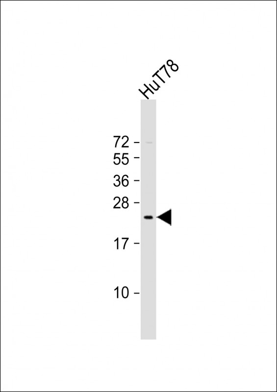
| Target/Specificity | This BATF3 antibody is generated from a rabbit immunized with a KLH conjugated synthetic peptide between 58-89 amino acids from the Central region of human BATF3. |
| Dilution | WB~~1:2000 E~~Use at an assay dependent concentration. |
| Format | Purified polyclonal antibody supplied in PBS with 0.09% (W/V) sodium azide. This antibody is purified through a protein A column, followed by peptide affinity purification. |
| Storage | Maintain refrigerated at 2-8°C for up to 2 weeks. For long term storage store at -20°C in small aliquots to prevent freeze-thaw cycles. |
| Precautions | BATF3 Antibody (Center) is for research use only and not for use in diagnostic or therapeutic procedures. |
| Name | BATF3 |
|---|---|
| Synonyms | SNFT |
| Function | AP-1 family transcription factor that controls the differentiation of CD8(+) thymic conventional dendritic cells in the immune system. Required for development of CD8-alpha(+) classical dendritic cells (cDCs) and related CD103(+) dendritic cells that cross- present antigens to CD8 T-cells and produce interleukin-12 (IL12) in response to pathogens (By similarity). Acts via the formation of a heterodimer with JUN family proteins that recognizes and binds DNA sequence 5'-TGA[CG]TCA-3' and regulates expression of target genes. |
| Cellular Location | Nucleus {ECO:0000255|PROSITE-ProRule:PRU00978, ECO:0000269|PubMed:12087103} |

Thousands of laboratories across the world have published research that depended on the performance of antibodies from Abcepta to advance their research. Check out links to articles that cite our products in major peer-reviewed journals, organized by research category.
info@abcepta.com, and receive a free "I Love Antibodies" mug.

Provided below are standard protocols that you may find useful for product applications.
Background
AP-1 family transcription factor that controls the differentiation of CD8(+) thymic conventional dendritic cells in the immune system. Required for development of CD8-alpha(+) classical dendritic cells (cDCs) and related CD103(+) dendritic cells that cross-present antigens to CD8 T-cells and produce interleukin-12 (IL12) in response to pathogens (By similarity). Acts via the formation of a heterodimer with JUN family proteins that recognizes and binds DNA sequence 5'-TGA[CG]TCA-3' and regulates expression of target genes.
References
Iacobelli M.,et al.J. Immunol. 165:860-868(2000).
Gregory S.G.,et al.Nature 441:315-321(2006).
Mural R.J.,et al.Submitted (SEP-2005) to the EMBL/GenBank/DDBJ databases.
Bower K.E.,et al.J. Biol. Chem. 277:34967-34977(2002).
Bower K.E.,et al.Oncogene 23:8805-8814(2004).
If you have used an Abcepta product and would like to share how it has performed, please click on the "Submit Review" button and provide the requested information. Our staff will examine and post your review and contact you if needed.
If you have any additional inquiries please email technical services at tech@abcepta.com.
Ordering Information
Other Products
Shipping Information
