


 Foundational characteristics of cancer include proliferation, angiogenesis, migration, evasion of apoptosis, and cellular immortality. Find key markers for these cellular processes and antibodies to detect them.
 The SUMOplot™ Analysis Program predicts and scores sumoylation sites in your protein. SUMOylation is a post-translational modification involved in various cellular processes, such as nuclear-cytosolic transport, transcriptional regulation, apoptosis, protein stability, response to stress, and progression through the cell cycle.
 The Autophagy Receptor Motif Plotter predicts and scores autophagy receptor binding sites in your protein. Identifying proteins connected to this pathway is critical to understanding the role of autophagy in physiological as well as pathological processes such as development, differentiation, neurodegenerative diseases, stress, infection, and cancer.
Epsin2 Antibody (N-term)
Purified Rabbit Polyclonal Antibody (Pab)
- SPECIFICATION
- CITATIONS: 1
- PROTOCOLS
- BACKGROUND

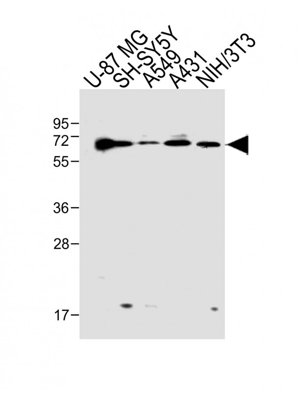
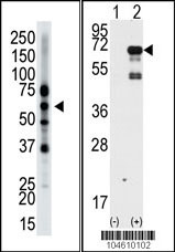
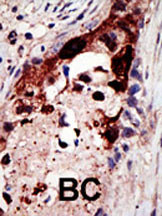
Application
| WB, IHC-P, E |
|---|---|
| Primary Accession | O95208 |
| Other Accession | Q9Z1Z3, Q8CHU3, O88339, Q80VP1, Q9Y6I3, Q8IYW4 |
| Reactivity | Human, Mouse |
| Predicted | Rat |
| Host | Rabbit |
| Clonality | Polyclonal |
| Isotype | Rabbit IgG |
| Calculated MW | 68482 Da |
| Antigen Region | 6-36 aa |
| Gene ID | 22905 |
|---|---|
| Other Names | Epsin-2, EPS-15-interacting protein 2, EPN2, KIAA1065 |
| Target/Specificity | This Epsin2 antibody is generated from rabbits immunized with a KLH conjugated synthetic peptide between 6-36 amino acids from the N-terminal region of human Epsin2. |
| Dilution | WB~~1:1000 IHC-P~~1:50~100 E~~Use at an assay dependent concentration. |
| Format | Purified polyclonal antibody supplied in PBS with 0.09% (W/V) sodium azide. This antibody is purified through a protein A column, followed by peptide affinity purification. |
| Storage | Maintain refrigerated at 2-8°C for up to 2 weeks. For long term storage store at -20°C in small aliquots to prevent freeze-thaw cycles. |
| Precautions | Epsin2 Antibody (N-term) is for research use only and not for use in diagnostic or therapeutic procedures. |
| Name | EPN2 |
|---|---|
| Synonyms | KIAA1065 |
| Function | Plays a role in the formation of clathrin-coated invaginations and endocytosis. |
| Cellular Location | Cytoplasm. Cytoplasmic vesicle, clathrin-coated vesicle. Note=In punctate structures throughout the cell, associated with clathrin-coated vesicles, and particularly concentrated in the region of the Golgi complex |
| Tissue Location | Highest expression is found in brain. Detected at lower levels in lung and liver. |

Provided below are standard protocols that you may find useful for product applications.
Background
Epsin2 plays a role in the formation of clathrin-coated invaginations and endocytosis. It binds to EPS15 (via the NPF repeat domain), as well as to AP-2 and clathrin (via the DPW repeat domain). The protein resides in the cytoplasm, in punctate structures throughout the cell, associated with clathrin-coated vesicles, and is particularly concentrated in the region of the Golgi complex. Highest expression is found in brain, with lower levels detected in lung and liver.
References
Ota, T., et al., Nat. Genet. 36(1):40-45 (2004).
Kikuno, R., et al., DNA Res. 6(3):197-205 (1999).
Rosenthal, J.A., et al., J. Biol. Chem. 274(48):33959-33965 (1999).
If you have used an Abcepta product and would like to share how it has performed, please click on the "Submit Review" button and provide the requested information. Our staff will examine and post your review and contact you if needed.
If you have any additional inquiries please email technical services at tech@abcepta.com.
Ordering Information
Other Products
Shipping Information
