


 Foundational characteristics of cancer include proliferation, angiogenesis, migration, evasion of apoptosis, and cellular immortality. Find key markers for these cellular processes and antibodies to detect them.
 The SUMOplot™ Analysis Program predicts and scores sumoylation sites in your protein. SUMOylation is a post-translational modification involved in various cellular processes, such as nuclear-cytosolic transport, transcriptional regulation, apoptosis, protein stability, response to stress, and progression through the cell cycle.
 The Autophagy Receptor Motif Plotter predicts and scores autophagy receptor binding sites in your protein. Identifying proteins connected to this pathway is critical to understanding the role of autophagy in physiological as well as pathological processes such as development, differentiation, neurodegenerative diseases, stress, infection, and cancer.
FXC1 Antibody (N-term)
Purified Rabbit Polyclonal Antibody (Pab)
- SPECIFICATION
- CITATIONS
- PROTOCOLS
- BACKGROUND

Application
| WB, E |
|---|---|
| Primary Accession | Q9Y5J6 |
| Other Accession | Q3SZW4, Q9WV96, Q5RDJ0, Q9R1B1 |
| Reactivity | Human, Rat |
| Predicted | Bovine, Mouse |
| Host | Rabbit |
| Clonality | polyclonal |
| Isotype | Rabbit IgG |
| Calculated MW | 11586 Da |
| Gene ID | 26515 |
|---|---|
| Other Names | Mitochondrial import inner membrane translocase subunit Tim10 B, Fracture callus protein 1, FxC1, Mitochondrial import inner membrane translocase subunit Tim9 B, TIMM10B, Tim10b, TIMM10B, FXC1, TIM9B, TIMM9B |
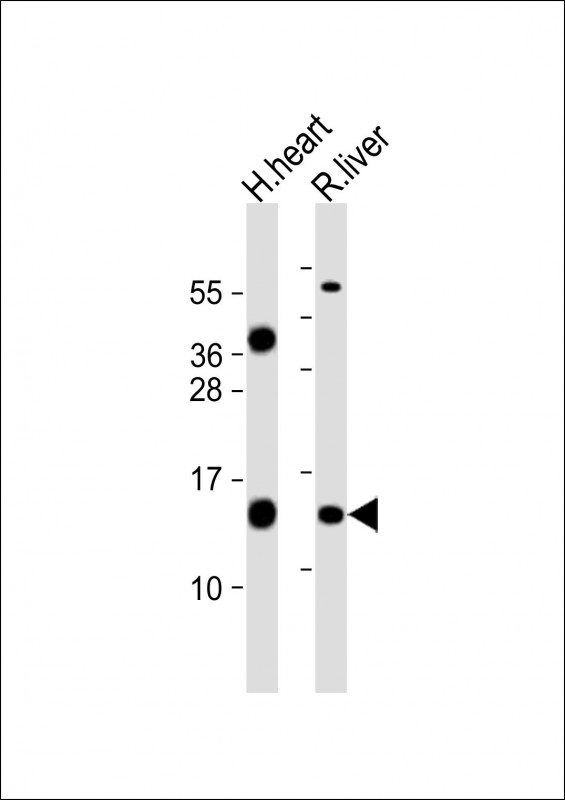
| Target/Specificity | This FXC1 antibody is generated from a rabbit immunized with a KLH conjugated synthetic peptide between 15-48 amino acids from the N-terminal region of human FXC1. |
| Dilution | WB~~1:2000 E~~Use at an assay dependent concentration. |
| Format | Purified polyclonal antibody supplied in PBS with 0.09% (W/V) sodium azide. This antibody is purified through a protein A column, followed by peptide affinity purification. |
| Storage | Maintain refrigerated at 2-8°C for up to 2 weeks. For long term storage store at -20°C in small aliquots to prevent freeze-thaw cycles. |
| Precautions | FXC1 Antibody (N-term) is for research use only and not for use in diagnostic or therapeutic procedures. |
| Name | TIMM10B |
|---|---|
| Synonyms | FXC1, TIM9B, TIMM9B |
| Function | Component of the TIM22 complex, a complex that mediates the import and insertion of multi-pass transmembrane proteins into the mitochondrial inner membrane. The TIM22 complex forms a twin-pore translocase that uses the membrane potential as the external driving force. In the TIM22 complex, it may act as a docking point for the soluble 70 kDa complex that guides the target proteins in transit through the aqueous mitochondrial intermembrane space. |
| Cellular Location | Mitochondrion inner membrane; Peripheral membrane protein |
| Tissue Location | Ubiquitous, with highest expression in heart, kidney, liver and skeletal muscle. |

Thousands of laboratories across the world have published research that depended on the performance of antibodies from Abcepta to advance their research. Check out links to articles that cite our products in major peer-reviewed journals, organized by research category.
info@abcepta.com, and receive a free "I Love Antibodies" mug.

Provided below are standard protocols that you may find useful for product applications.
Background
Component of the TIM22 complex, a complex that mediates the import and insertion of multi-pass transmembrane proteins into the mitochondrial inner membrane. The TIM22 complex forms a twin- pore translocase that uses the membrane potential as the external driving force. In the TIM22 complex, it may act as a docking point for the soluble 70 kDa complex that guides the target proteins in transit through the aqueous mitochondrial intermembrane space.
References
Jin H.,et al.Genomics 61:259-267(1999).
Bauer M.F.,et al.FEBS Lett. 464:41-47(1999).
Peng Y.,et al.Submitted (SEP-1999) to the EMBL/GenBank/DDBJ databases.
Rothbauer U.,et al.J. Biol. Chem. 276:37327-37334(2001).
Muehlenbein N.,et al.J. Biol. Chem. 279:13540-13546(2004).
If you have used an Abcepta product and would like to share how it has performed, please click on the "Submit Review" button and provide the requested information. Our staff will examine and post your review and contact you if needed.
If you have any additional inquiries please email technical services at tech@abcepta.com.
Ordering Information
Other Products
Shipping Information
