


 Foundational characteristics of cancer include proliferation, angiogenesis, migration, evasion of apoptosis, and cellular immortality. Find key markers for these cellular processes and antibodies to detect them.
 The SUMOplot™ Analysis Program predicts and scores sumoylation sites in your protein. SUMOylation is a post-translational modification involved in various cellular processes, such as nuclear-cytosolic transport, transcriptional regulation, apoptosis, protein stability, response to stress, and progression through the cell cycle.
 The Autophagy Receptor Motif Plotter predicts and scores autophagy receptor binding sites in your protein. Identifying proteins connected to this pathway is critical to understanding the role of autophagy in physiological as well as pathological processes such as development, differentiation, neurodegenerative diseases, stress, infection, and cancer.
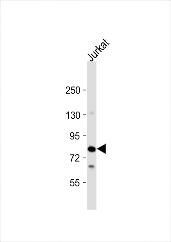
LCP2 Antibody (N-Term)
Purified Rabbit Polyclonal Antibody (Pab)
- SPECIFICATION
- CITATIONS
- PROTOCOLS
- BACKGROUND

Application
| WB, E |
|---|---|
| Primary Accession | Q13094 |
| Reactivity | Human |
| Host | Rabbit |
| Clonality | polyclonal |
| Isotype | Rabbit IgG |
| Calculated MW | 60188 Da |
| Gene ID | 3937 |
|---|---|
| Other Names | Lymphocyte cytosolic protein 2, SH2 domain-containing leukocyte protein of 76 kDa, SLP-76 tyrosine phosphoprotein, SLP76, LCP2 |
| Target/Specificity | This LCP2 antibody is generated from a rabbit immunized with a KLH conjugated synthetic peptide between 46-80 amino acids from human LCP2. |
| Dilution | WB~~1:2000 E~~Use at an assay dependent concentration. |
| Format | Purified polyclonal antibody supplied in PBS with 0.09% (W/V) sodium azide. This antibody is purified through a protein A column, followed by peptide affinity purification. |
| Storage | Maintain refrigerated at 2-8°C for up to 2 weeks. For long term storage store at -20°C in small aliquots to prevent freeze-thaw cycles. |
| Precautions | LCP2 Antibody (N-Term) is for research use only and not for use in diagnostic or therapeutic procedures. |
| Name | LCP2 |
|---|---|
| Function | Adapter protein primarily involved in signaling pathways within T-cells, as well as other immune cells such as platelets, mast cells, and natural killer (NK) cells (PubMed:11313406, PubMed:33159816). Plays a crucial role for transducing signal from the T-cell receptor (TCR) after antigen recognition leading to T-cell activation. Mechanistically, once phosphorylated by the kinase ZAP70, mediates interactions with the guanine-nucleotide exchange factor VAV1, the adapter protein NCK and the kinase ITK (PubMed:8673706, PubMed:8702662). In turn, stimulates the activation of PKC-theta/PRKCQ and NF-kappa-B transcriptional activity in response to CD3 and CD28 costimulation (PubMed:11313406). Also plays an essential role in AGER- induced signaling pathways including p38 MAPK and ERK1/2 activation leading to cytokine release and pro-inflammatory responses (PubMed:33436632). |
| Cellular Location | Cytoplasm. |
| Tissue Location | Highly expressed in spleen, thymus and peripheral blood leukocytes. Highly expressed also in T-cell and monocytic cell lines, expressed at lower level in B-cell lines. Not detected in fibroblast or neuroblastoma cell lines |

Thousands of laboratories across the world have published research that depended on the performance of antibodies from Abcepta to advance their research. Check out links to articles that cite our products in major peer-reviewed journals, organized by research category.
info@abcepta.com, and receive a free "I Love Antibodies" mug.

Provided below are standard protocols that you may find useful for product applications.
Background
Involved in T-cell antigen receptor mediated signaling.
References
Jackman J.K.,et al.J. Biol. Chem. 270:7029-7032(1995).
Kalnine N.,et al.Submitted (MAY-2003) to the EMBL/GenBank/DDBJ databases.
Ota T.,et al.Nat. Genet. 36:40-45(2004).
Mural R.J.,et al.Submitted (SEP-2005) to the EMBL/GenBank/DDBJ databases.
Moog-Lutz C.,et al.J. Biol. Chem. 276:22375-22381(2001).
If you have used an Abcepta product and would like to share how it has performed, please click on the "Submit Review" button and provide the requested information. Our staff will examine and post your review and contact you if needed.
If you have any additional inquiries please email technical services at tech@abcepta.com.
Ordering Information
Other Products
Shipping Information
