


 Foundational characteristics of cancer include proliferation, angiogenesis, migration, evasion of apoptosis, and cellular immortality. Find key markers for these cellular processes and antibodies to detect them.
 The SUMOplot™ Analysis Program predicts and scores sumoylation sites in your protein. SUMOylation is a post-translational modification involved in various cellular processes, such as nuclear-cytosolic transport, transcriptional regulation, apoptosis, protein stability, response to stress, and progression through the cell cycle.
 The Autophagy Receptor Motif Plotter predicts and scores autophagy receptor binding sites in your protein. Identifying proteins connected to this pathway is critical to understanding the role of autophagy in physiological as well as pathological processes such as development, differentiation, neurodegenerative diseases, stress, infection, and cancer.
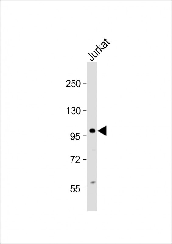
KIAA1524 Antibody (Center)
Purified Rabbit Polyclonal Antibody (Pab)
- SPECIFICATION
- CITATIONS
- PROTOCOLS
- BACKGROUND

Application
| WB, E |
|---|---|
| Primary Accession | Q8TCG1 |
| Reactivity | Human |
| Host | Rabbit |
| Clonality | polyclonal |
| Isotype | Rabbit IgG |
| Calculated MW | 102185 Da |
| Gene ID | 57650 |
|---|---|
| Other Names | Protein CIP2A, Cancerous inhibitor of PP2A, p90 autoantigen, KIAA1524, CIP2A |
| Target/Specificity | This KIAA1524 antibody is generated from a rabbit immunized with a KLH conjugated synthetic peptide between 537-567 amino acids from the Central region of human KIAA1524. |
| Dilution | WB~~1:2000 E~~Use at an assay dependent concentration. |
| Format | Purified polyclonal antibody supplied in PBS with 0.09% (W/V) sodium azide. This antibody is purified through a protein A column, followed by peptide affinity purification. |
| Storage | Maintain refrigerated at 2-8°C for up to 2 weeks. For long term storage store at -20°C in small aliquots to prevent freeze-thaw cycles. |
| Precautions | KIAA1524 Antibody (Center) is for research use only and not for use in diagnostic or therapeutic procedures. |
| Name | CIP2A {ECO:0000303|PubMed:17632056, ECO:0000312|HGNC:HGNC:29302} |
|---|---|
| Function | Acts as an inhibitor of protein phosphatase PP2A (PubMed:17632056). Promotes anchorage-independent cell growth and tumor formation by preventing dephosphorylation of MYC, thereby stabilizing MYC in human malignancies (PubMed:17632056). Together with TOPBP1, plays an essential role in the response to genome instability generated by the presence of acentric chromosome fragments derived from shattered chromosomes within micronuclei (PubMed:35121901, PubMed:35842428, PubMed:37165191, PubMed:37316668). Micronuclei, which are frequently found in cancer cells, consist of chromatin surrounded by their own nuclear membrane: following breakdown of the micronuclear envelope, a process associated with chromothripsis, the CIP2A-TOPBP1 complex tethers chromosome fragments during mitosis to ensure clustered segregation of the fragments to a single daughter cell nucleus, facilitating re-ligation with limited chromosome scattering and loss (PubMed:37165191, PubMed:37316668). |
| Cellular Location | Cytoplasm. Chromosome. Note=Predominantly localizes within the cytoplasm (PubMed:35842428). Localizes to broken chromosomes within micronuclei during interphase and following chromothripsis (PubMed:37165191, PubMed:37316668). Localization to broken chromosomes is mainly independent of MDC1 (PubMed:35121901, PubMed:37165191) |
| Tissue Location | Expressed at low levels in most of the tissues. Overexpressed in head-and-neck squamous cell carcinomas (HNSCC) Present in liver cancer cells (at protein level) |

Thousands of laboratories across the world have published research that depended on the performance of antibodies from Abcepta to advance their research. Check out links to articles that cite our products in major peer-reviewed journals, organized by research category.
info@abcepta.com, and receive a free "I Love Antibodies" mug.

Provided below are standard protocols that you may find useful for product applications.
Background
Oncoprotein that inhibits PP2A and stabilizes MYC in human malignancies. Promotes anchorage-independent cell growth and tumor formation.
References
Soo Hoo L.,et al.Oncogene 21:5006-5015(2002).
Nagase T.,et al.DNA Res. 7:143-150(2000).
Bechtel S.,et al.BMC Genomics 8:399-399(2007).
Ota T.,et al.Nat. Genet. 36:40-45(2004).
Shi F.D.,et al.Prostate 63:252-258(2005).
If you have used an Abcepta product and would like to share how it has performed, please click on the "Submit Review" button and provide the requested information. Our staff will examine and post your review and contact you if needed.
If you have any additional inquiries please email technical services at tech@abcepta.com.
Ordering Information
Shipping Information
