


 Foundational characteristics of cancer include proliferation, angiogenesis, migration, evasion of apoptosis, and cellular immortality. Find key markers for these cellular processes and antibodies to detect them.
 The SUMOplot™ Analysis Program predicts and scores sumoylation sites in your protein. SUMOylation is a post-translational modification involved in various cellular processes, such as nuclear-cytosolic transport, transcriptional regulation, apoptosis, protein stability, response to stress, and progression through the cell cycle.
 The Autophagy Receptor Motif Plotter predicts and scores autophagy receptor binding sites in your protein. Identifying proteins connected to this pathway is critical to understanding the role of autophagy in physiological as well as pathological processes such as development, differentiation, neurodegenerative diseases, stress, infection, and cancer.
MGEA5 Antibody (N-Term)
Purified Rabbit Polyclonal Antibody (Pab)
- SPECIFICATION
- CITATIONS
- PROTOCOLS
- BACKGROUND

Application
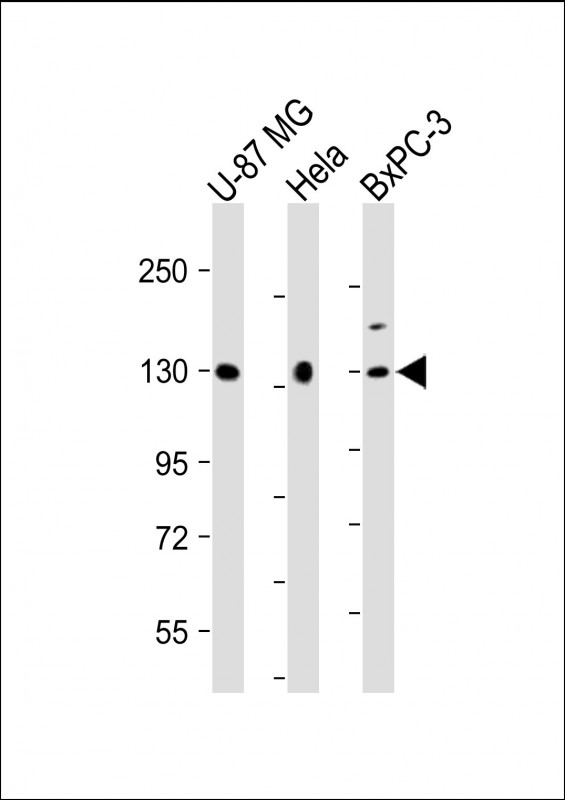
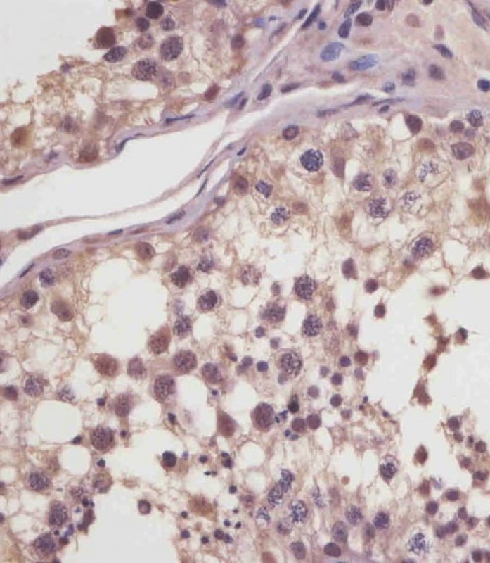
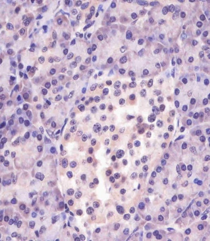
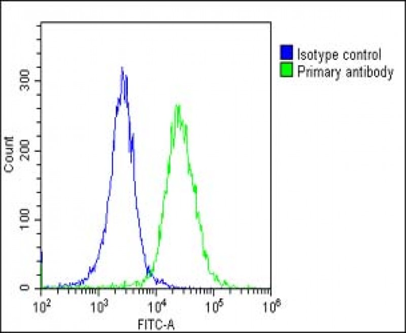
| WB, FC, IHC-P, E |
|---|---|
| Primary Accession | O60502 |
| Other Accession | Q9EQQ9, Q8VIJ5 |
| Reactivity | Human |
| Predicted | Mouse, Rat |
| Host | Rabbit |
| Clonality | polyclonal |
| Isotype | Rabbit IgG |
| Calculated MW | 102915 Da |
| Gene ID | 10724 |
|---|---|
| Other Names | Protein O-GlcNAcase, OGA, 3.2.1.169, Beta-N-acetylglucosaminidase, 3.2.1.-, Beta-N-acetylhexosaminidase, Beta-hexosaminidase, Meningioma-expressed antigen 5, N-acetyl-beta-D-glucosaminidase, N-acetyl-beta-glucosaminidase, Nuclear cytoplasmic O-GlcNAcase and acetyltransferase, NCOAT, MGEA5, HEXC, KIAA0679, MEA5 |
| Target/Specificity | This MGEA5 antibody is generated from a rabbit immunized with a KLH conjugated synthetic peptide between 236-269 amino acids from the human region of human MGEA5. |
| Dilution | WB~~1:2000 FC~~1:25 IHC-P~~1:25 E~~Use at an assay dependent concentration. |
| Format | Purified polyclonal antibody supplied in PBS with 0.09% (W/V) sodium azide. This antibody is purified through a protein A column, followed by peptide affinity purification. |
| Storage | Maintain refrigerated at 2-8°C for up to 2 weeks. For long term storage store at -20°C in small aliquots to prevent freeze-thaw cycles. |
| Precautions | MGEA5 Antibody (N-Term) is for research use only and not for use in diagnostic or therapeutic procedures. |
| Name | OGA {ECO:0000303|PubMed:20863279, ECO:0000312|HGNC:HGNC:7056} |
|---|---|
| Function | [Isoform 1]: Cleaves GlcNAc but not GalNAc from O- glycosylated proteins (PubMed:11148210, PubMed:11788610, PubMed:20673219, PubMed:22365600, PubMed:24088714, PubMed:28939839, PubMed:37962578). Deglycosylates a large and diverse number of proteins, such as CRYAB, ELK1, GSDMD, LMNB1 and TAB1 (PubMed:28939839, PubMed:37962578). Can use p-nitrophenyl-beta-GlcNAc and 4- methylumbelliferone-GlcNAc as substrates but not p-nitrophenyl-beta- GalNAc or p-nitrophenyl-alpha-GlcNAc (in vitro) (PubMed:20673219). Does not bind acetyl-CoA and does not have histone acetyltransferase activity (PubMed:24088714). |
| Cellular Location | [Isoform 3]: Nucleus |
| Tissue Location | Ubiquitous. Shows highest expression in the brain, placenta and pancreas. |

Thousands of laboratories across the world have published research that depended on the performance of antibodies from Abcepta to advance their research. Check out links to articles that cite our products in major peer-reviewed journals, organized by research category.
info@abcepta.com, and receive a free "I Love Antibodies" mug.

Provided below are standard protocols that you may find useful for product applications.
Background
Isoform 1: Cleaves GlcNAc but not GalNAc from O- glycosylated proteins. Can use p-nitrophenyl-beta-GlcNAc and 4- methylumbelliferone-GlcNAc as substrates but not p-nitrophenyl- beta-GalNAc or p-nitrophenyl-alpha-GlcNAc (in vitro) (PubMed:11148210). Does not bind acetyl-CoA and does not have histone acetyltransferase activity (PubMed:24088714).
References
Heckel D.,et al.Hum. Mol. Genet. 7:1859-1872(1998).
Comtesse N.,et al.Biochem. Biophys. Res. Commun. 283:634-640(2001).
Gao Y.,et al.J. Biol. Chem. 276:9838-9845(2001).
Ishikawa K.,et al.DNA Res. 5:169-176(1998).
Nakajima D.,et al.DNA Res. 9:99-106(2002).
If you have used an Abcepta product and would like to share how it has performed, please click on the "Submit Review" button and provide the requested information. Our staff will examine and post your review and contact you if needed.
If you have any additional inquiries please email technical services at tech@abcepta.com.
Ordering Information
Other Products
Shipping Information
