


 Foundational characteristics of cancer include proliferation, angiogenesis, migration, evasion of apoptosis, and cellular immortality. Find key markers for these cellular processes and antibodies to detect them.
 The SUMOplot™ Analysis Program predicts and scores sumoylation sites in your protein. SUMOylation is a post-translational modification involved in various cellular processes, such as nuclear-cytosolic transport, transcriptional regulation, apoptosis, protein stability, response to stress, and progression through the cell cycle.
 The Autophagy Receptor Motif Plotter predicts and scores autophagy receptor binding sites in your protein. Identifying proteins connected to this pathway is critical to understanding the role of autophagy in physiological as well as pathological processes such as development, differentiation, neurodegenerative diseases, stress, infection, and cancer.
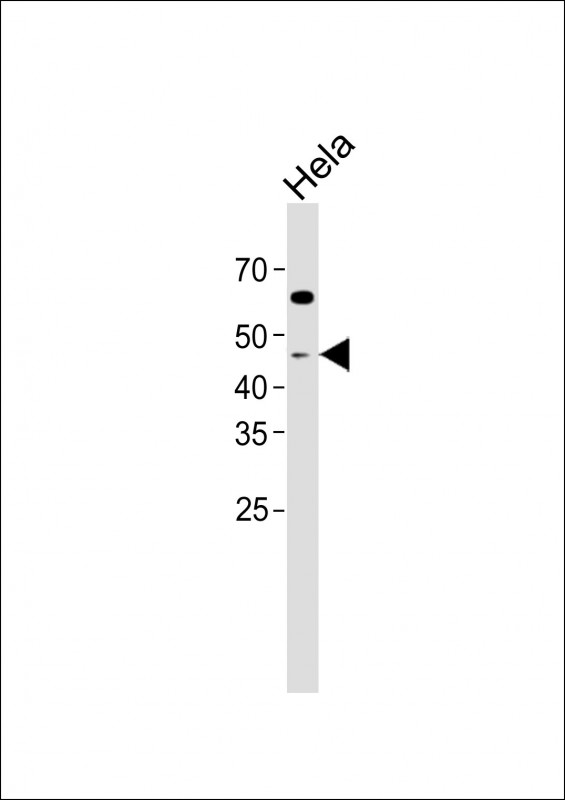
anti-Caspase 9 Antibody
Purified Rabbit Polyclonal Antibody (Pab)
- SPECIFICATION
- CITATIONS
- PROTOCOLS
- BACKGROUND

Application
| WB, E |
|---|---|
| Primary Accession | P55211 |
| Reactivity | Human |
| Host | Rabbit |
| Clonality | polyclonal |
| Isotype | Rabbit Ig |
| Calculated MW | 46281 Da |
| Gene ID | 842 |
|---|---|
| Other Names | Caspase-9, CASP-9, 3.4.22.62, Apoptotic protease Mch-6, Apoptotic protease-activating factor 3, APAF-3, ICE-like apoptotic protease 6, ICE-LAP6, Caspase-9 subunit p35, Caspase-9 subunit p10, CASP9, MCH6 |
| Target/Specificity | This anti-Caspase 9 antibody is generated from a rabbit immunized with a KLH conjugated synthetic peptide between amino acids from the human region of human anti-Caspase 9. |
| Dilution | WB~~1:2000 E~~Use at an assay dependent concentration. |
| Format | Purified polyclonal antibody supplied in PBS with 0.09% (W/V) sodium azide. This antibody is purified through a protein A column, followed by peptide affinity purification. |
| Storage | Maintain refrigerated at 2-8°C for up to 2 weeks. For long term storage store at -20°C in small aliquots to prevent freeze-thaw cycles. |
| Precautions | anti-Caspase 9 Antibody is for research use only and not for use in diagnostic or therapeutic procedures. |
| Name | CASP9 |
|---|---|
| Synonyms | MCH6 |
| Function | Involved in the activation cascade of caspases responsible for apoptosis execution. Binding of caspase-9 to Apaf-1 leads to activation of the protease which then cleaves and activates effector caspases caspase-3 (CASP3) or caspase-7 (CASP7). Promotes DNA damage- induced apoptosis in a ABL1/c-Abl-dependent manner. Proteolytically cleaves poly(ADP-ribose) polymerase (PARP). Cleaves BIRC6 following inhibition of BIRC6-caspase binding by DIABLO/SMAC (PubMed:36758105, PubMed:36758106). |
| Tissue Location | Ubiquitous, with highest expression in the heart, moderate expression in liver, skeletal muscle, and pancreas. Low levels in all other tissues. Within the heart, specifically expressed in myocytes. |

Thousands of laboratories across the world have published research that depended on the performance of antibodies from Abcepta to advance their research. Check out links to articles that cite our products in major peer-reviewed journals, organized by research category.
info@abcepta.com, and receive a free "I Love Antibodies" mug.

Provided below are standard protocols that you may find useful for product applications.
Background
Involved in the activation cascade of caspases responsible for apoptosis execution. Binding of caspase-9 to Apaf-1 leads to activation of the protease which then cleaves and activates effector caspases caspase-3 (CASP3) or caspase-7 (CASP7). Promotes DNA damage- induced apoptosis in a ABL1/c-Abl-dependent manner. Proteolytically cleaves poly(ADP-ribose) polymerase (PARP).
References
Duan H.,et al.J. Biol. Chem. 271:16720-16724(1996).
Srinivasula S.M.,et al.J. Biol. Chem. 271:27099-27106(1996).
Hadano S.,et al.Mamm. Genome 10:757-760(1999).
Srinivasula S.M.,et al.Cancer Res. 59:999-1002(1999).
Izawa M.,et al.Submitted (JUN-1998) to the EMBL/GenBank/DDBJ databases.
If you have used an Abcepta product and would like to share how it has performed, please click on the "Submit Review" button and provide the requested information. Our staff will examine and post your review and contact you if needed.
If you have any additional inquiries please email technical services at tech@abcepta.com.
Ordering Information
Other Products
Shipping Information
