


 Foundational characteristics of cancer include proliferation, angiogenesis, migration, evasion of apoptosis, and cellular immortality. Find key markers for these cellular processes and antibodies to detect them.
 The SUMOplot™ Analysis Program predicts and scores sumoylation sites in your protein. SUMOylation is a post-translational modification involved in various cellular processes, such as nuclear-cytosolic transport, transcriptional regulation, apoptosis, protein stability, response to stress, and progression through the cell cycle.
 The Autophagy Receptor Motif Plotter predicts and scores autophagy receptor binding sites in your protein. Identifying proteins connected to this pathway is critical to understanding the role of autophagy in physiological as well as pathological processes such as development, differentiation, neurodegenerative diseases, stress, infection, and cancer.
Ribophorin 2 (RPN2) Antibody (N-term)
Purified Rabbit Polyclonal Antibody (Pab)
- SPECIFICATION
- CITATIONS: 1
- PROTOCOLS
- BACKGROUND

Application
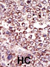
| IHC-P, E |
|---|---|
| Primary Accession | P04844 |
| Other Accession | P25235, Q9GL01, Q3SZI6, NP_002942 |
| Reactivity | Human |
| Predicted | Bovine, Pig, Rat |
| Host | Rabbit |
| Clonality | Polyclonal |
| Isotype | Rabbit IgG |
| Calculated MW | 69284 Da |
| Antigen Region | 16-46 aa |
| Gene ID | 6185 |
|---|---|
| Other Names | Dolichyl-diphosphooligosaccharide--protein glycosyltransferase subunit 2, Dolichyl-diphosphooligosaccharide--protein glycosyltransferase 63 kDa subunit, RIBIIR, Ribophorin II, RPN-II, Ribophorin-2, RPN2 |
| Target/Specificity | This Ribophorin 2 (RPN2) antibody is generated from rabbits immunized with a KLH conjugated synthetic peptide between 16-46 amino acids from the N-terminal region of human Ribophorin 2 (RPN2). |
| Dilution | IHC-P~~1:50~100 E~~Use at an assay dependent concentration. |
| Format | Purified polyclonal antibody supplied in PBS with 0.09% (W/V) sodium azide. This antibody is prepared by Saturated Ammonium Sulfate (SAS) precipitation followed by dialysis against PBS. |
| Storage | Maintain refrigerated at 2-8°C for up to 2 weeks. For long term storage store at -20°C in small aliquots to prevent freeze-thaw cycles. |
| Precautions | Ribophorin 2 (RPN2) Antibody (N-term) is for research use only and not for use in diagnostic or therapeutic procedures. |
| Name | RPN2 (HGNC:10382) |
|---|---|
| Function | Subunit of the oligosaccharyl transferase (OST) complex that catalyzes the initial transfer of a defined glycan (Glc(3)Man(9)GlcNAc(2) in eukaryotes) from the lipid carrier dolichol- pyrophosphate to an asparagine residue within an Asn-X-Ser/Thr consensus motif in nascent polypeptide chains, the first step in protein N-glycosylation (PubMed:31831667). N-glycosylation occurs cotranslationally and the complex associates with the Sec61 complex at the channel-forming translocon complex that mediates protein translocation across the endoplasmic reticulum (ER). All subunits are required for a maximal enzyme activity. |
| Cellular Location | Endoplasmic reticulum {ECO:0000250|UniProtKB:F1PCT7}. Endoplasmic reticulum membrane; Multi- pass membrane protein |
| Tissue Location | Expressed in all tissues tested. |

Provided below are standard protocols that you may find useful for product applications.
Background
RNP2 a type I integral membrane protein found only in the rough endoplasmic reticulum. The encoded protein is part of an N-oligosaccharyl transferase complex that links high mannose oligosaccharides to asparagine residues found in the Asn-X-Ser/Thr consensus motif of nascent polypeptide chains. This protein is similar in sequence to the yeast oligosaccharyl transferase subunit SWP1.
References
Kelleher, D.J., et al., Mol. Cell 12(1):101-111 (2003). Fu, J., et al., J. Biol. Chem. 275(6):3984-3990 (2000). Loffler, C., et al., Hum. Genet. 87(2):221-222 (1991). Crimaudo, C., et al., EMBO J. 6(1):75-82 (1987). Stoffel, M., et al., Hum. Mol. Genet. 1 (8), 656 (1992).
If you have used an Abcepta product and would like to share how it has performed, please click on the "Submit Review" button and provide the requested information. Our staff will examine and post your review and contact you if needed.
If you have any additional inquiries please email technical services at tech@abcepta.com.
Ordering Information
Other Products
Shipping Information
