


 Foundational characteristics of cancer include proliferation, angiogenesis, migration, evasion of apoptosis, and cellular immortality. Find key markers for these cellular processes and antibodies to detect them.
 The SUMOplot™ Analysis Program predicts and scores sumoylation sites in your protein. SUMOylation is a post-translational modification involved in various cellular processes, such as nuclear-cytosolic transport, transcriptional regulation, apoptosis, protein stability, response to stress, and progression through the cell cycle.
 The Autophagy Receptor Motif Plotter predicts and scores autophagy receptor binding sites in your protein. Identifying proteins connected to this pathway is critical to understanding the role of autophagy in physiological as well as pathological processes such as development, differentiation, neurodegenerative diseases, stress, infection, and cancer.
INDO Antibody (Center)
Purified Rabbit Polyclonal Antibody (Pab)

- SPECIFICATION
- CITATIONS
- PROTOCOLS
- BACKGROUND

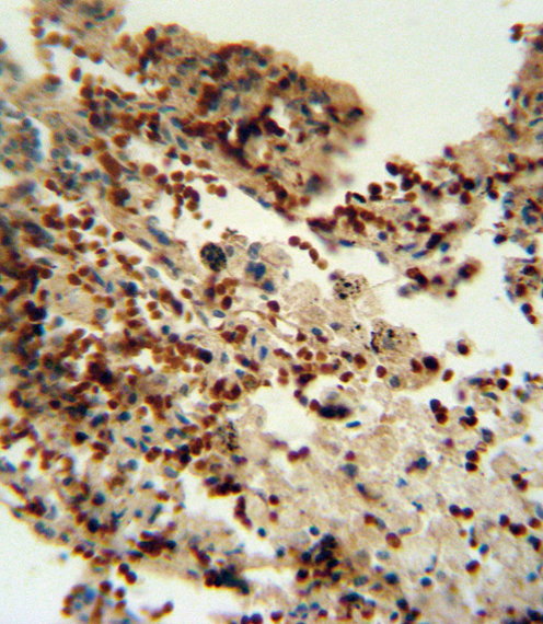
Application
| IHC-P, WB, E |
|---|---|
| Primary Accession | P14902 |
| Other Accession | NP_002155.1 |
| Reactivity | Human, Mouse |
| Host | Rabbit |
| Clonality | Polyclonal |
| Isotype | Rabbit IgG |
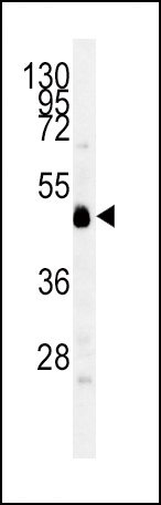
| Calculated MW | 45326 Da |
| Antigen Region | 79-105 aa |
| Gene ID | 3620 |
|---|---|
| Other Names | Indoleamine 2, 3-dioxygenase 1, IDO-1, Indoleamine-pyrrole 2, 3-dioxygenase, IDO1, IDO, INDO |
| Target/Specificity | This INDO antibody is generated from rabbits immunized with a KLH conjugated synthetic peptide between 79-105 amino acids from the Central region of human INDO. |
| Dilution | IHC-P~~1:10~50 WB~~1:1000 E~~Use at an assay dependent concentration. |
| Format | Purified polyclonal antibody supplied in PBS with 0.09% (W/V) sodium azide. This antibody is prepared by Saturated Ammonium Sulfate (SAS) precipitation followed by dialysis against PBS. |
| Storage | Maintain refrigerated at 2-8°C for up to 2 weeks. For long term storage store at -20°C in small aliquots to prevent freeze-thaw cycles. |
| Precautions | INDO Antibody (Center) is for research use only and not for use in diagnostic or therapeutic procedures. |
| Name | IDO1 (HGNC:6059) |
|---|---|
| Synonyms | IDO, INDO |
| Function | Catalyzes the first and rate limiting step of the catabolism of the essential amino acid tryptophan along the kynurenine pathway (PubMed:17671174). Involved in the peripheral immune tolerance, contributing to maintain homeostasis by preventing autoimmunity or immunopathology that would result from uncontrolled and overreacting immune responses (PubMed:25691885). Tryptophan shortage inhibits T lymphocytes division and accumulation of tryptophan catabolites induces T-cell apoptosis and differentiation of regulatory T-cells (PubMed:25691885). Acts as a suppressor of anti-tumor immunity (PubMed:14502282, PubMed:23103127, PubMed:25157255, PubMed:25691885). Limits the growth of intracellular pathogens by depriving tryptophan (PubMed:25691885). Protects the fetus from maternal immune rejection (PubMed:25691885). |
| Cellular Location | Cytoplasm, cytosol {ECO:0000250|UniProtKB:P28776, ECO:0000303|PubMed:25691885} |
| Tissue Location | Expressed in mature dendritic cells located in lymphoid organs (including lymph nodes, spleen, tonsils, Peyers's patches, the gut lamina propria, and the thymic medulla), in some epithelial cells of the female genital tract, as well as in endothelial cells of term placenta and in lung parenchyma (PubMed:25691885). Weakly or not expressed in most normal tissues, but mostly inducible in most tissues (PubMed:25691885). Expressed in more than 50% of tumors, either by tumoral, stromal, or endothelial cells (expression in tumor is associated with a worse clinical outcome) (PubMed:18418598). Not overexpressed in tumor-draining lymph nodes (PubMed:25691885, PubMed:26155395). |
Research Areas
Citations (0)

Thousands of laboratories across the world have published research that depended on the performance of antibodies from Abcepta to advance their research. Check out links to articles that cite our products in major peer-reviewed journals, organized by research category.
Submit your citation using an Abcepta antibody to
info@abcepta.com, and receive a free "I Love Antibodies" mug.
info@abcepta.com, and receive a free "I Love Antibodies" mug.

Application Protocols
Provided below are standard protocols that you may find useful for product applications.
Abcepta welcomes feedback from its customers.
If you have used an Abcepta product and would like to share how it has performed, please click on the "Submit Review" button and provide the requested information. Our staff will examine and post your review and contact you if needed.
If you have any additional inquiries please email technical services at tech@abcepta.com.
$ 150.00
$ 385.00
Cat# AP2728c
Ordering Information
United States
AlbaniaAustraliaAustriaBelgiumBosnia & HerzegovinaBrazilBulgariaCanadaCentral AmericaChinaCroatiaCyprusCzech RepublicDenmarkEstoniaFinlandFranceGermanyGreeceHong KongHungaryIcelandIndiaIndonesiaIrelandIsraelItalyJapanLatviaLithuaniaLuxembourgMacedoniaMalaysiaMaltaMexicoNetherlandsNew ZealandNorwayPakistanPolandPortugalRomaniaSerbiaSingaporeSlovakiaSloveniaSouth AfricaSouth KoreaSpainSwedenSwitzerlandTaiwanTurkeyUnited KingdomUnited StatesVietnamWorldwideOthers
USA Headquarters
(888) 735-7227 / (858) 622-0099 or (858) 875-1900
Other Products
Shipping Information
Domestic orders (in stock items)
Shipped out the same day. Orders placed after 1 PM (PST) will ship out the next business day.
International orders
Contact your local distributors
