


 Foundational characteristics of cancer include proliferation, angiogenesis, migration, evasion of apoptosis, and cellular immortality. Find key markers for these cellular processes and antibodies to detect them.
 The SUMOplot™ Analysis Program predicts and scores sumoylation sites in your protein. SUMOylation is a post-translational modification involved in various cellular processes, such as nuclear-cytosolic transport, transcriptional regulation, apoptosis, protein stability, response to stress, and progression through the cell cycle.
 The Autophagy Receptor Motif Plotter predicts and scores autophagy receptor binding sites in your protein. Identifying proteins connected to this pathway is critical to understanding the role of autophagy in physiological as well as pathological processes such as development, differentiation, neurodegenerative diseases, stress, infection, and cancer.
Phospho-TNFR(S274) Antibody
Affinity Purified Rabbit Polyclonal Antibody (Pab)
- SPECIFICATION
- CITATIONS
- PROTOCOLS
- BACKGROUND

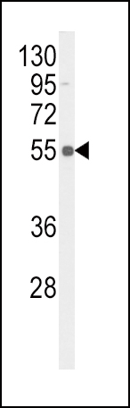
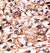
Application
| WB, IHC-P, E |
|---|---|
| Primary Accession | P19438 |
| Reactivity | Human |
| Host | Rabbit |
| Clonality | Polyclonal |
| Isotype | Rabbit IgG |
| Calculated MW | 50495 Da |
| Gene ID | 7132 |
|---|---|
| Other Names | Tumor necrosis factor receptor superfamily member 1A, Tumor necrosis factor receptor 1, TNF-R1, Tumor necrosis factor receptor type I, TNF-RI, TNFR-I, p55, p60, CD120a, Tumor necrosis factor receptor superfamily member 1A, membrane form, Tumor necrosis factor-binding protein 1, TBPI, TNFRSF1A, TNFAR, TNFR1 |
| Target/Specificity | This TNFR Antibody is generated from rabbits immunized with a KLH conjugated synthetic phosphopeptide corresponding to amino acid residues surrounding S274 of human TNFR. |
| Dilution | WB~~1:1000 IHC-P~~1:50~100 E~~Use at an assay dependent concentration. |
| Format | Purified polyclonal antibody supplied in PBS with 0.09% (W/V) sodium azide. This antibody is purified through a protein A column, followed by peptide affinity purification. |
| Storage | Maintain refrigerated at 2-8°C for up to 2 weeks. For long term storage store at -20°C in small aliquots to prevent freeze-thaw cycles. |
| Precautions | Phospho-TNFR(S274) Antibody is for research use only and not for use in diagnostic or therapeutic procedures. |
| Name | TNFRSF1A |
|---|---|
| Synonyms | TNFAR, TNFR1 |
| Function | Receptor for TNFSF2/TNF-alpha and homotrimeric TNFSF1/lymphotoxin-alpha. The adapter molecule FADD recruits caspase-8 to the activated receptor. The resulting death-inducing signaling complex (DISC) performs caspase-8 proteolytic activation which initiates the subsequent cascade of caspases (aspartate-specific cysteine proteases) mediating apoptosis. Contributes to the induction of non-cytocidal TNF effects including anti-viral state and activation of the acid sphingomyelinase. |
| Cellular Location | Cell membrane; Single-pass type I membrane protein Golgi apparatus membrane; Single-pass type I membrane protein. Secreted. Note=A secreted form is produced through proteolytic processing |

Thousands of laboratories across the world have published research that depended on the performance of antibodies from Abcepta to advance their research. Check out links to articles that cite our products in major peer-reviewed journals, organized by research category.
info@abcepta.com, and receive a free "I Love Antibodies" mug.

Provided below are standard protocols that you may find useful for product applications.
Background
A member of the TNF-receptor superfamily, this protein is one of the major receptors for the tumor necrosis factor-alpha. This receptor can activate NF-kappaB, mediate apoptosis, and function as a regulator of inflammation. Antiapoptotic protein BCL2-associated athanogene 4 (BAG4/SODD) and adaptor proteins TRADD and TRAF2 have been shown to interact with this receptor, and thus play regulatory roles in the signal transduction mediated by the receptor. Germline mutations of the extracellular domains of this receptor were found to be associated with the autosomal dominant periodic fever syndrome. The impaired receptor clearance is thought to be a mechanism of the disease.
References
Kuo, N.W., et al., Invest. Ophthalmol. Vis. Sci. 46(5):1565-1571 (2005). Siebert, S., et al., Arthritis Rheum. 52(4):1287-1292 (2005). Spahr, L., et al., J. Hepatol. 41(2):229-234 (2004). Wang, W.H., et al., Mol. Cell. Biol. 24(23):10352-10365 (2004). Tashiro, H., et al., Transpl. Int. 17(10):626-633 (2004).
If you have used an Abcepta product and would like to share how it has performed, please click on the "Submit Review" button and provide the requested information. Our staff will examine and post your review and contact you if needed.
If you have any additional inquiries please email technical services at tech@abcepta.com.
Ordering Information
Other Products
Shipping Information
