


 Foundational characteristics of cancer include proliferation, angiogenesis, migration, evasion of apoptosis, and cellular immortality. Find key markers for these cellular processes and antibodies to detect them.
 The SUMOplot™ Analysis Program predicts and scores sumoylation sites in your protein. SUMOylation is a post-translational modification involved in various cellular processes, such as nuclear-cytosolic transport, transcriptional regulation, apoptosis, protein stability, response to stress, and progression through the cell cycle.
 The Autophagy Receptor Motif Plotter predicts and scores autophagy receptor binding sites in your protein. Identifying proteins connected to this pathway is critical to understanding the role of autophagy in physiological as well as pathological processes such as development, differentiation, neurodegenerative diseases, stress, infection, and cancer.
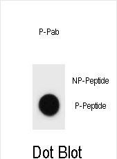
Phospho-CCNB3(T280) Antibody
Affinity Purified Rabbit Polyclonal Antibody (Pab)
- SPECIFICATION
- CITATIONS
- PROTOCOLS
- BACKGROUND

Application
| DB, E |
|---|---|
| Primary Accession | Q8WWL7 |
| Other Accession | NP_149020.2 |
| Reactivity | Human |
| Host | Rabbit |
| Clonality | Polyclonal |
| Isotype | Rabbit IgG |
| Calculated MW | 157916 Da |
| Gene ID | 85417 |
|---|---|
| Other Names | G2/mitotic-specific cyclin-B3, CCNB3, CYCB3 |
| Target/Specificity | This CCNB3 Antibody is generated from rabbits immunized with a KLH conjugated synthetic phosphopeptide corresponding to amino acid residues surrounding T280 of human CCNB3. |
| Dilution | DB~~1:500 E~~Use at an assay dependent concentration. |
| Format | Purified polyclonal antibody supplied in PBS with 0.09% (W/V) sodium azide. This antibody is purified through a protein A column, followed by peptide affinity purification. |
| Storage | Maintain refrigerated at 2-8°C for up to 2 weeks. For long term storage store at -20°C in small aliquots to prevent freeze-thaw cycles. |
| Precautions | Phospho-CCNB3(T280) Antibody is for research use only and not for use in diagnostic or therapeutic procedures. |
| Name | CCNB3 |
|---|---|
| Synonyms | CYCB3 |
| Function | Cyclins are positive regulatory subunits of the cyclin- dependent kinases (CDKs), and thereby play an essential role in the control of the cell cycle, notably via their destruction during cell division. Its tissue specificity suggest that it may be required during early meiotic prophase I. |
| Cellular Location | Nucleus. |
| Tissue Location | Testis specific. In testis, it is expressed in developing germ cells, but not in Leydig cells. Weakly or not expressed in other tissues. |

Thousands of laboratories across the world have published research that depended on the performance of antibodies from Abcepta to advance their research. Check out links to articles that cite our products in major peer-reviewed journals, organized by research category.
info@abcepta.com, and receive a free "I Love Antibodies" mug.

Provided below are standard protocols that you may find useful for product applications.
Background
The protein encoded by this gene belongs to the highly conserved cyclin family, whose members are characterized by a dramatic periodicity in protein abundance through the cell cycle. Cyclins function as regulators of CDK kinases. Different cyclins exhibit distinct expression and degradation patterns which contribute to the temporal coordination of each mitotic event. Studies of similar genes in chick and Drosophila suggest that this cyclin may associate with CDC2 and CDK2 kinases, and be required for proper spindle reorganization and restoration of the interphase nucleus. Two transcript variants encoding different isoforms have been found for this gene.
References
Cheng, J., et al. Science 308(5725):1149-1154(2005)
Nguyen, T.B., et al. J. Biol. Chem. 277(44):41960-41969(2002)
Hrimech, M., et al. EMBO J. 21 (14), 3918 (2002) :
Lozano, J.C., et al. Biochem. Biophys. Res. Commun. 291(2):406-413(2002)
Mouland, A.J., et al. Virology 292(2):321-330(2002)
If you have used an Abcepta product and would like to share how it has performed, please click on the "Submit Review" button and provide the requested information. Our staff will examine and post your review and contact you if needed.
If you have any additional inquiries please email technical services at tech@abcepta.com.
Ordering Information
Other Products
Shipping Information
